


-

M. Opitz
deepeye Medical, Munich, Germany M. Opitz
deepeye Medical, Munich, GermanyECS01 – deepeye Medical
Seeing More, In Less Time. deepeye® TPS is the world’s first AI assistant for predictive retinal Therapy Planning Support and automated documentation.
deepeye Medical GmbH (DPY) is a heathtech company headquartered in Munich and provides clinicians with an AI assistant for retinal therapies. Our mission is to empower ophthalmologists to see more clinical detail while spending less time on manual analysis and documentation. Our flagship solution, deepeye® TPS (Therapy Planning Support), acts like an “AI-voice-scribe” – but for imaging, transforming complex OCT histories into actionable, one-glance clinical insights.
Seeing More: Clinical Depth & Predictive Power
Beyond Segmentation: While traditional OCT AI focuses on biomarkers, deepeye® TPS provides a disease activity assessment and a comprehensive 12-month therapy response prognosis for anti-VEGF. This allows clinicians to move from reactive to proactive care, answering the patient’s most vital question: “What is my personal therapy outlook?”
Objective Trend Analysis: The system “reads” the OCTs of the last 1-2 years to visualize disease activity progression for therapy success as well as changes to a treatment need prognosis. By identifying subtle “Good vs. Bad Responder” patterns, it goes beyond biomarkers with its traffic-light charts. It has shown to positively influence therapy decisions (e.g. earlier interval shorterning) and thus can support clinicians in keeping patients on the optimal to stable vision.
The “deepeye Effect”: By providing objective data for interval planning, the system supports physicians in evidence-led patient retention and might improve long-term visual stabilization, compared to traditional real-world outcomes.In Less Time: Unmatched Operational Efficiency
3x Faster Review: deepeye® TPS delivers up to 60% time saving per OCT analysis and documentation. For high-volume clinics, this translates to gaining up to one hour of surgery time every day, enabling less stress & manageable workloads for the team and increasing patient throughput.
90% Less Typing: The AI analysis automatically provides a standardized text summaries for EMRs. This can drastically reduce the burden of manual documentation, ensuring standardized OCT analysis records for quality assurance by payors with minimal effort.
Seamless Workflow: DPY is device-agnostic (Topcon, Zeiss, Heidelberg, Optopol …) and works with all major image management platforms (Heyex, FORUM, Harmony etc.) as well as EMRs.Key Outcome Goals
Clinical Excellence: Need-based therapy planning through predictive, data-driven interval planning and efficient & effective patient education.
Human-in-the-Loop Safety: Enhanced therapy decision confidence thanks to assistive AI (that does not replace a doctor’s decision!), fulfilling the requirements of the EU AI Act and ophthalmic ethical AI guidelines (BVA/DOG/EURETINA).
Economic Sustainability: Boost practice ROI by increasing efficiency and reducing possible risks, e.g. incomplete documentation, incorrect billing and quality assurance findings by payors.Company website: https://www.deepeye-medical.com/
Supporting documents

-

J. Berestka
Ophthalmology, Northwest Eye, Minneapolis, United States J. Berestka
Ophthalmology, Northwest Eye, Minneapolis, United StatesECS02 – Lightfield Medical
Lightfield Medical: Solving ophthalmology’s missing link — autonomous slit-lamp imaging that technicians can capture and doctors can trust
The slit lamp has anchored anterior segment examination for nearly a century, yet the exam remains ephemeral — visible only to the clinician present, gone when the patient leaves. Demand for ophthalmic care is projected to rise 24% by 2035 while the ophthalmologist workforce shrinks. The only solution is delegation, and delegation requires a device that a technician can operate without specialized skill. That device has not existed — until now.
Conventional slit lamp cameras require a skilled operator and produce unstandardized images across nine independent degrees of freedom. Nothing is comparable across visits, and meaningful technician delegation is not possible.
Lightfield Medical designed Captura from first principles. A dual tunable liquid lens system autofocuses the full anterior segment automatically. A programmable digital light engine generates any illumination pattern on demand. A non-parfocal optical design captures the complete exam with a single moving component. A technician presses one button and a standardized, navigable digital record is produced in seconds.
The programmable illumination unlocks imaging impossible on a conventional slit lamp. Compound slits combine two illumination types in one frame, cutting acquisition time and storage costs by half. A miniaturized slit prism with 780nm illumination produces the world’s first full-pupil retroillumination image — no white bar artifact, no dilation — enabling routine technician screening for Fuchs’ dystrophy, ABMD, and cataract. Annular sclerotic scatter, in development, will be the first of many further novel modalities the platform is expected to unlock.
The Captura Viewer iOS app lets a reviewing physician navigate the captured exam by gesture from any location. Routine follow-up visits can be handled by technicians at remote sites with asynchronous physician review. One physician can cover multiple retail optical locations remotely — a critical capability given that roughly half of Americans currently skip their recommended annual eye exam due to a shortage of on-site optometrists.
Because every Captura image is captured under identical software-defined conditions, the dataset is reproducible across every device in the field — the prerequisite for reliable AI training.
Lightfield Medical operates on a DaaS subscription model across a global market of more than 80,000 ophthalmology practices. The company holds granted patents in the US and Europe with prosecution underway in eight additional jurisdictions — hundreds of claims covering the core technology and designed to block competitive workarounds. The strategic path targets acquisition by a major ophthalmic platform, and conversations with relevant parties are underway.
Company website: www.lightfieldmedical.com
Supporting documents
-

N. Katchinskiy
Electrical and Computer Engineering, University of Alberta, Edmonton, Canada N. Katchinskiy
Electrical and Computer Engineering, University of Alberta, Edmonton, CanadaECS03 – PulseMedica
PulseMedica SVO-Tx Image-Guided Femtosecond Laser System
PulseMedica is pioneering ophthalmic laser surgery with the world’s first image-guided femtosecond laser platform designed to image, diagnose, and treat vitreoretinal disease. Our lead clinical application targets vitreous opacities, or eye floaters, which represent a major unmet need in ophthalmology and are one of the most common patient complaints. This includes post-PVD and post-cataract patients who remain symptomatic despite otherwise successful eye care, and who cannot access, are not candidates for, or do not want vitrectomy or off-label YAG vitreolysis.
Over 50% of adults experience floaters, and 10-25% suffer moderate-to-severe visual impairment and distraction affecting daily function. Imaging and treating floaters is technologically difficult because floaters are small, mobile, translucent collagen-based structures within the vitreous, requiring objective imaging, real-time tracking, treatment planning, and precise laser delivery near sensitive anatomy.
The dominant approach is “wait and see,” which leaves symptomatic patients without meaningful treatment. Vitrectomy can remove floaters but is invasive and carries risks such as retinal tears, endophthalmitis, and early-onset cataract formation. Off-label YAG vitreolysis is manual, imprecise, not FDA-approved for floater treatment, and lacks consistent effectiveness and purpose-built safety features. Current care also lacks objective diagnostic tools, treatment planning capabilities, and a standardized referral pathway.
PulseMedica’s platform system solves these shortcomings by integrating imaging, planning, and treatment delivery into one physician-supervised workflow. The system combines OCT/SLO imaging, a patient docking interface for eye stabilization, AI-based floater tracking, automated treatment volume execution, and femtosecond laser photoionization. Unlike vitrectomy or off-label YAG vitreolysis, PulseMedica’s approach is purpose-built for floater imaging and treatment, non-invasive, and designed to reduce risk from human error and subjective judgment. By precisely delivering laser volumes while tracking floater motion and avoiding heat-based tissue disruption, the platform is designed to enable objective diagnosis and effective treatment through a fast, predictable, repeatable, office-based procedure.
Company website: www.pulsemedica.com
Supporting documents
-

D. May
Ophthalmology, HelpMeSee. Inc, Jersey city, United States D. May
Ophthalmology, HelpMeSee. Inc, Jersey city, United StatesECS04 – Ophthalmology, HelpMeSee. Inc
Scaling Cataract Surgery Training Through High-Fidelity Simulation: The HelpMeSee Competency-Based Approach to Building Global Surgical Capacity
Cataract remains one of the world’s leading causes of preventable blindness, yet access to quality cataract surgery is limited in many regions by a shortage of trained surgeons and inconsistent opportunities for hands-on training. Traditional surgical education relies heavily on live patient exposure, variable case availability, mentor-dependent teaching, and gradual skill acquisition under high-pressure clinical conditions. These constraints can lengthen the learning curve and limit the ability to repeatedly practice critical steps or rare complications before entering the operating room.
HelpMeSee addresses this training bottleneck through a simulation-based surgical education system designed specifically for cataract care. Founded on principles adapted from aviation simulation, HelpMeSee combines high-fidelity virtual reality, haptic feedback that mimics tactile tissue interaction, structured curriculum, instructor-led training, and real-time objective performance feedback. This approach allows cataract specialists to practice essential procedural steps repeatedly in a risk-free environment. It also builds hand-eye coordination, confidence and provides trainees standardized guidance before transitioning to live surgery.
The HelpMeSee training portfolio includes Manual Small Incision Cataract Surgery (MSICS) training, Phacoemulsification training, and supplementary modules covering complex or complication-focused scenarios such as Posterior Capsular rupture Management, Zonular Dialysis Management, Secondary IOL Techniques, Small Pupil Management, and Suturing. By enabling unlimited repetition, consistent instruction, measurable progress, and exposure to simulated complications, the system supports competency-based learning rather than relying solely on time-in-training or chance case exposure.The HelpMeSee global model is designed for scalability through training centers, partner institutions, and locally integrated pathways. The organization’s mission is to restore vision to people living with severe visual impairment and blindness due to cataract worldwide, while its strategy focuses on innovative, scalable, technology-driven surgical simulation training. At ESCRS iNovation Day, HelpMeSee will present how simulation can help strengthen cataract surgical education and improve readiness for live surgery. This improves patient safety and supports the growth of sustainable surgical capacity, especially important in regions with the greatest need.
Company website: www.helpmesee.org
Supporting documents
-

D. Rodrigues
Acting CEO and Board of Directors, VisiRose, Inc. - President and Vice Chairman, Board of Directors, Provectus Biopharmaceuticals, Inc., Knoxville, United States D. Rodrigues
Acting CEO and Board of Directors, VisiRose, Inc. - President and Vice Chairman, Board of Directors, Provectus Biopharmaceuticals, Inc., Knoxville, United StatesECS05 – VisiRose, Inc.
Rose Bengal Photodynamic Antimicrobial Therapy (RB-PDAT): A Resistance-Proof, Broad-Spectrum Treatment for Infectious Keratitis and Corneal Disease — Developed at Bascom Palmer Eye Institute
VisiRose, Inc.
VisiRose is a clinical-stage ophthalmic biotechnology company advancing Rose Bengal Photodynamic Antimicrobial Therapy (RB-PDAT), a non-invasive, light-activated treatment for infectious keratitis developed at Bascom Palmer Eye Institute, the #1-ranked U.S. eye hospital.
The Technology
RB-PDAT is a drug-device combination treatment that pairs rose bengal — a synthetic small molecule with 100+ years of safe ophthalmic use — with targeted green-light activation to destroy pathogens via reactive oxygen species. The mechanism is physical, not biological: no resistance has been documented across any pathogen class. The proprietary active pharmaceutical ingredient is manufactured under Current Good Manufacturing Practice using a Quality by Design synthesis process developed in accordance with International Council on Harmonisation guidelines to meet chemistry, manufacturing, and control regulatory requirements. One mechanism. Indifferent to pathogen class. Hard to resist.
The Market Opportunity
Infectious keratitis affects 5 to 10 million patients annually and is a leading cause of corneal blindness worldwide. When antimicrobials fail and drug-resistant bacterial, fungal, parasitic, and viral strains are making that increasingly likely, the fallback is corneal transplant surgery. RB-PDAT eliminates that fallback. The addressable global market for rose bengal-based ophthalmic therapies exceeds $20 billion, spanning antimicrobial resistance-concern markets in the U.S. and Europe, high-incidence markets across Asia, Latin America, and Africa, and adjacent ophthalmic indications enabled by the molecule’s intrinsic mechanisms.
The Evidence Base
36 peer-reviewed publications covering in vitro, in vivo, clinical, and mechanistic work; 280+ patients treated across six institutions in four countries on three continents. The REAGIR Phase 3 RCT (NCT05110001) — 330 patients, double-masked, sham-controlled — delivered the first positive signal against natamycin as standard of care in nearly 50 years: directionally favorable results in the Fusarium subgroup, the trial’s largest and most powered cohort.
Key Outcome Goals
Regulatory approval in the U.S., Europe, and key international markets for Fusarium fungal infectious keratitis. Protocol optimization informed by REAGIR subgroup findings. Indication expansion into infectious scleritis, corneal ectasia, keratoconus, and floppy eyelid syndrome. A scalable, low-cost delivery model deployable across academic and community ophthalmology settings globally.
RB-PDAT is a platform with the potential to redefine the standard of care for corneal infection — reducing preventable blindness, displacing costly surgery, and outpacing microbial resistance.
Company website: www.visirose.com
Supporting documents
-

M. Mrochen
Management, Allotex, Milano, Italy - Management, IROC Science to Innovation, Zug - Management, Pallas Kliniken, Olten, Switzerland M. Mrochen
Management, Allotex, Milano, Italy - Management, IROC Science to Innovation, Zug - Management, Pallas Kliniken, Olten, SwitzerlandECS06 – Allotex
Allotex pioneers tissue-addition vision correction for presbyopia. By adding natural, sterile corneal collagen, it restores the eye’s range of vision while preserving optical quality and future treatment options—already commercially launched and advancing on the U.S. regulatory pathway.
Company non-confidential summary: The global “presbyopia gap” represents a large, underserved segment—approximately 30% of the population aged 40–60 with emmetropia or low ametropia—who want near vision restoration without sacrificing distance vision or committing to irreversible surgery.
Allotex addresses this high-value segment with a differentiated, biologic solution: a sterile, ethically sourced human collagen allograft, optically shaped with micron-level precision. This additive technology preserves corneal integrity and maintains all future treatment options—positioning it as a category-defining innovation. With accelerating commercial growth in Europe, Canada, and other regions, an active U.S. FDA clinical pathway, and a focused APAC expansion strategy, Allotex is building a scalable global platform and establishing Tissue Addition Technology as a new standard in refractive surgery.Company website: eu.allotex.com/
Supporting documents
-
I. Pallikaris
PRES LTD - Laboratory of Optics and Vision, University of Crete, HERAKLION, Greece I. Pallikaris
PRES LTD - Laboratory of Optics and Vision, University of Crete, HERAKLION, GreeceECS07 – PRES LTD
Customised keratorefractive surface or lenticule extraction procedure using specialised molded material prior to femtosecond laser incision(s)
Our company is developing a novel approach to corneal refractive surgery designed to overcome key limitations of existing laser-based procedures such as LASIK, PRK, and KLEX. While these techniques have significantly improved visual outcomes, they remain limited in their ability to fully correct corneal irregularities and higher-order aberrations, especially in complex or abnormal corneas.
Our proprietary technology enables customised keratorefractive surface or lenticule extraction procedures without requiring modifications to femtosecond laser platforms or specialised software. The innovation involves applying a biocompatible material to the corneal surface prior to laser docking. This material is molded and polymerised to create a perfectly smooth anterior corneal curvature that serves as a reference surface for the first laser incision.
By temporarily masking corneal irregularities, the method enables the creation of a spherical interface during the initial lamellar cut. Following removal of the molded material, surface extraction procedures eliminate anterior corneal irregularities, leaving a smooth spherical surface that corrects refractive error. In lenticule extraction procedures, a second planar incision captures and corrects native corneal irregularities. The resulting lenticule has adual-surface functionality: one surface optimised for spherical refractive correction and the other tailored to correct complex optical distortions.
This approach replicates the optical quality of rigid contact lenses, enabling a level of precision and regularity not possible with current refractive procedures. It also simplifies the surgical workflow by relying on one or two planar laser incisions and removes dependence on imperfect wavefront or topography-guided measurements.
Our objectives are to improve visual quality by reducing higher-order aberrations and enhancing contrast sensitivity, particularly in low-light conditions; expand the treatable patient population to include irregular corneas and higher refractive errors; and reduce variability and the learning curve associated with existing procedures through a more predictable and reproducible technique. Compatibility with current laser systems lowers adoption barriers and supports rapid integration into clinical practices.
With a validated proof of concept ex vivo and ongoing process optimisation, the company is advancing toward clinical development and regulatory approval. Our long-term vision is to establish this technology as a new standard of care in refractive surgery, capturing a meaningful share of a growing global market while delivering optimised outcomes for patients worldwide.
Company website: www.pres-ltd.com
Supporting documents
-

Y. Yahav
RevitalVision , Modi'in , Israel Y. Yahav
RevitalVision , Modi'in , IsraelECS08 – RevitalVision
Improving Vision Beyond the Eyes with Patient-Specific Visual Stimulation
About our company:
RevitalVision is a SaaS-based digital health startup headquartered in Israel at an initial growth stage.
We offer a unique solution to millions with poor or insufficient vision whose needs are unmet by existing vision treatments. Our non-invasive technology enhances eyesight neurologically. We utilize brain science and proprietary algorithms to visually stimulate the brain’s visual cortex to improve vision. Our technology has been clinically validated to enhance vision outcomes with long-lasting results in several eye diseases and vision conditions, with a significant impact on vision and quality of life, with an average vision improvement of more than 2 ETDRS lines and 100% improvement in contrast sensitivity. Our product — a home-based vision training app — operates directly from a standard web browser on any personal computer, without the need for any special hardware. Our customers include: 1) Eyecare professionals (B2B) who prescribe the home-based treatment with our software for specific conditions such as amblyopia (lazy eye) and for patients with eye diseases, and 2) Consumers (B2C) seeking wellness and post-surgery solutions beyond available treatments.
Our technology is the only FDA-cleared product with an efficacy claim to improve vision in amblyopia (lazy eye) in patients aged 9 or older and is CE marked with a broad intent for use — to improve vision above age 9.
In 2025, RevitalVision demonstrated efficacy and safety in seven randomized controlled clinical studies, three of which were published in leading peer-reviewed ophthalmology journals (AJO, BJO, and LAJO). The studies investigated RevitalVision efficacy in the following indications: Keratoconus, nystagmus, and post-cataract surgery with multifocal IOL’s and the long-term efficacy (5 years) on adults with amblyopia.
Company website: www.revitalvision.com
Supporting documents
-

M. Webb
Epion Therapeutics, Burlington, United States M. Webb
Epion Therapeutics, Burlington, United StatesECS09 – Epion Therapeutics
Vision Ahead of the Curve: EpiSmart Epi-on CXL for Keratoconus
Epion is a late-stage (Phase 3) biotech company developing a non-invasive treatment for keratoconus, a progressive corneal disease that can cause vision loss and blindness.
Epion’s new therapy, the EpiSmart™ Epithelium-On Cross-Linking System, is a non-invasive drug-device combination designed to replace current, highly invasive therapies for keratoconus. EpiSmart is designed to treat keratoconus across a wide spectrum of disease severity, including on diagnosis, through simultaneous bilateral, non-invasive treatment. With an epithelium-on approach without chemical or physical disruption of the epithelium, EpiSmart may significantly reduce patient discomfort and recovery time, allowing for rapid return to daily activities and increasing access to vision-preserving care for millions of patients globally.
Funded by the AXA Global Health Fund and over 50 cornea specialists, Epion completed enrollment in its Phase 3 trials in April 2025 and expects to have results from its Phase 3 trial by August 2026. Epion is currently seeking investment in its Series B venture capital-priced round to accelerate FDA filing and prepare for commercialization.
The safety and efficacy of EpiSmart was investigated in a recently concluded 2,258-subject Phase 2 trial. Results of Epion recent Phase 2 trial, the largest study to-date of corneal cross-linking as a treatment for keratoconus, indicate improvements in visual acuity after EpiSmart treatment at levels similar to currently available treatments, but with a substantial enhancement in safety and comfort compared to the current standard of care.
Company website: www.epiontx.com
Supporting documents
-

M. Gomez
2EYESVISION, Madrid, Spain M. Gomez
2EYESVISION, Madrid, SpainECS10 – 2EYESVISION
Experience Vision Before Cataract and Presbyopia Surgery
2EyesVision is a Madrid-based ophthalmic technology company developing and commercializing SimVis Gekko2, a wearable, programmable visual simulator that lets patients experience in real time how different intraocular lenses could affect their vision before cataract or presbyopia surgery. Unlike screen-based or purely illustrative tools, SimVis Gekko2 provides a real-world binocular visual experience, helping surgeons and patients make more informed and personalized decisions in a setting where lens choice is otherwise irreversible and often based on explanation rather than direct experience.
The company’s goal is to improve preoperative decision-making, increase patient satisfaction, support adoption of premium IOL procedures, and establish a new standard of care in cataract and presbyopia surgery. The technology is built on proprietary CSIC-originated know-how, has been validated in more than 500 patients with 10,000+ simulations and 10+ clinical trials, and is backed by 30+ scientific publications and 5 patent families.
Company website: www.2eyesvision.com
Supporting documents
-

X. Dong
Thondar, Shenzhen, China X. Dong
Thondar, Shenzhen, ChinaECS11 – Thondar
Refraction Topography: A New Dimension for Myopia Assessment and Clinical Decision-Making Through Retinal Defocus Mapping
Refraction topography (RT) is a novel peripheral refraction measurement tool that can acquire peripheral refraction data across a 53° field of view using rapid focal scanning techniques.
RT develops a refraction algorithm on fundus images at various focusing statuses. Unlike conventional techniques for peripheral refraction measurement, RT requires the subject to stare at a stationary fixation target. The refraction algorithm calculates the focus measure for multiple images at the Point of Interest and formulates them into a focus profile. Its accuracy and repeatability have been recently validated in model eyes, adults, and children. RT makes RPRE visible and measurable, enabling earlier and more precise myopia management, as well as personalized treatment strategies.
Based on clinical application experience in the Chinese market, the use of Refraction Topography can be broadly categorized into four key areas:
1. Early Myopia Risk Assessment
By analyzing defocus patterns beyond 30° eccentricity and in the temporal retina, early changes associated with axial elongation can be identified, enabling prediction of myopia progression.
2. Orthokeratology Lens Fitting Evaluation
By capturing images at the central, nasal, and temporal regions and reconstructing up to a 150° retinal profile, clinicians can assess the area, size, and position of myopic defocus. This allows earlier evaluation of treatment efficacy before detectable changes in axial length.
3. Personalized Lens Design
Based on individual retinal defocus profiles, customized freeform designs on the posterior surface of lenses can be applied to compensate and optimize defocus distribution, improving the effectiveness of myopia control.
4. New Insights for Refractive Surgery
Provides a new analytical dimension for preoperative examination; introducing peripheral defocus data can be used to adjust surgical strategies, increase myopic defocus design, help reduce postoperative regression, and improve surgical outcomes.
Company website: www.thondar.com
Supporting documents
-

A. Pallikaris
EYE PCR BV, Amsterdam, Netherlands - Laboratory of Optics and Vision, University of Crete, Heraklion, Greece A. Pallikaris
EYE PCR BV, Amsterdam, Netherlands - Laboratory of Optics and Vision, University of Crete, Heraklion, GreeceECS12 – EYE PCR BV
Novel open bag endocapsular device fixOflex – Moving forward to market commercialization and expansion of indications of use
EYE PCR BV is an ophthalmic medical technology company focused on advancing natural human lens replacement surgery through an innovative endocapsular device platform. Its flagship product, fixOflex, is a recently CE-marked implantable device designed to preserve the 3-dimensional shape and intracapsular volume of the capsular bag following lens replacement surgery. Unlike conventional cataract surgery, where the capsular bag progressively contracts after surgery, fixOflex mechanically stabilizes the capsule and creates a more physiological environment for intraocular lens (IOL) positioning and long-term performance.
The device is intended to address several unmet needs in modern lens replacement surgery. By maintaining capsular integrity and stability, fixOflex aims to reduce postoperative capsular contraction and fibrosis, thereby improving posterior capsule opacification (PCO) prevention, IOL centration and rotational stability, and refractive predictability. The device is also designed to support future IOL exchange or repositioning by preserving intracapsular space and surgical access over time.
The technology platform was developed through more than 15 years of translational research and clinical investigation. A recent published peer-reviewed clinical study demonstrated encouraging safety and efficacy outcomes, including reduced PCO incidence for at least 12 months. Based on these clinical findings, fixOflex recently received CE mark approval with indications for preserving capsular form and intracapsular space while reducing PCO for at least 12 months following surgery.
The company’s current objective is to expand the clinical evidence base and broaden the technology’s potential indications. Ongoing clinical studies are evaluating the long-term effects of fixOflex on PCO prevention, its application in premium IOL procedures, and its potential role in pediatric ophthalmology. Additional benchmark and laboratory investigations are further characterizing the biomechanical effects of the device on capsular bag anatomy and stability.
EYE PCR BV is also initiating a soft commercial launch in selected target markets, with broader expansion planned as manufacturing capacity scales. The technology is particularly relevant for the growing premium IOL market, where PCO prevention, lens positioning accuracy, and long-term stability are critical for optimal visual outcomes and patient satisfaction.
The company’s broader vision is to redefine the role of the capsular bag in lens surgery by transforming it from a passive postoperative structure into a stable and durable platform for premium optics and future intraocular therapies.
Through fixOflex, the company aims to establish a new standard in cataract and refractive lens surgery focused on long-term anatomical preservation, optical stability, surgical flexibility, and lifelong patient benefit.
Company website: www.eyepcr.com
Supporting documents
-

A. Cerviño
University of Valencia, Valencia, Spain A. Cerviño
University of Valencia, Valencia, SpainECS13 – PairedFocus
A New Non-Diffractive Approach to Obtain Near Vision in Presbyopia-Correcting Intraocular Lenses
Background / Unmet Need: Presbyopia-correcting intraocular lenses (IOLs) continue to require a compromise between near visual performance and overall optical quality. While extended depth of focus (EDOF) IOLs provide excellent distance and intermediate vision with reduced photic phenomena, near visual acuity often remains suboptimal. Conversely, trifocal IOLs improve near vision but are frequently associated with dysphotopsia and reduced contrast sensitivity. This ongoing trade-off highlights the need for alternative optical strategies that enhance near vision without sacrificing visual quality.
Innovation: PairedFocus represents a novel, non-diffractive, wavefront-shaping IOL concept designed to extend functional vision into the near range while preserving distance optical performance. Unlike conventional EDOF or diffractive multifocal designs, this approach redistributes optical energy to optimize near vision without introducing diffractive steps, potentially reducing the risk of photic phenomena.
Scientific Rationale and Early Evidence: Preclinical optical bench testing was conducted in accordance with ISO standards to assess through-focus modulation transfer function and simulated visual performance. The near-optimized design demonstrated preserved distance optical quality together with enhanced performance at vergences beyond +2.50 D, resulting in a dual-peak defocus profile. Both alone or combined binocularly with a companion non-diffractive EDOF IOL, the system produced a continuous defocus curve with predicted functional visual acuity across the full range from distance to near. These findings support a complementary binocular strategy, referred to as the Balanced Extended Vision Procedure (BEVPx).
Clinical Relevance and Development Status: This non-diffractive concept aims to bridge the gap between EDOF and trifocal IOLs, offering the potential for improved near vision while maintaining visual quality and minimizing optical side effects. Early clinical experience with this technology is currently being evaluated, with clinical data scheduled for presentation during the ESCRS Congress.
Conclusion: PairedFocus introduces a new optical approach to presbyopia correction that may represent an emerging category of non-diffractive IOLs. By addressing an important unmet clinical need, this technology has the potential to expand the current spectrum of presbyopia-correcting solutions available to cataract and refractive surgeons.
Company website: www.astvc.com
Supporting documents
-

S. O’Neil
CEO, ViaLase, Aliso Viejo, United States S. O’Neil
CEO, ViaLase, Aliso Viejo, United StatesECS14 – ViaLase
FLIGHT: A Novel, Non-Surgical, Image-Guided Femtosecond Laser Approach to Trabeculotomy for Glaucoma
ViaLase is developing a femtosecond laser platform designed specifically for glaucoma treatment. The FLIGHT procedure (Femtosecond Laser Image-Guided High-Precision Trabeculotomy) uses high-resolution OCT imaging and ultrafast laser pulses to create precise trabeculotomy channels through the cornea without incisions or implants.
The company is led by pioneers in femtosecond laser technology whose earlier work helped establish ultrafast lasers as a transformative platform for ophthalmic surgery.
The objective of this presentation is to introduce the technology and its underlying mechanism, review the clinical rationale for a non-incisional trabeculotomy approach, and discuss early clinical experience and potential implications for the future management of glaucoma.
Company website: www.vialase.com
Supporting documents
-

T. G. Qiu
Ophthalmic Therapeutic Innovation, Inc., Peabody, United States T. G. Qiu
Ophthalmic Therapeutic Innovation, Inc., Peabody, United StatesECS15 – Ophthalmic Therapeutic Innovation
Transform Glaucoma Patient Care via Matrix Metalloproteinase (MMP) pathway Based Therapy
Ophthalmic Therapeutic Innovation, Inc is a clinical stage biotech that develops a breakthrough therapy for treating glaucomatous neural degeneration via matrix metalloproteinase (MMP) pathway activation and tissue rejuvenation. OTI was founded in 2022 by Dr Tina Guanting Qiu, a retinal surgeon and stem cell biologist with decades of drug development experience in industry (GSK, Inotek Pharma, Astellas Pharma). Dr Qiu pioneers big data for IP and regulatory innovation, and made a breakthrough discovery of what causes glaucoma: the pressure sensing cell loss and sensor functional damage (irregularities) is the rate limiting root cause of vision loss in glaucoma. The company is at Seed stage financing with a late stage clinical asset, raised 250,000 pre-seed fund from glaucoma surgeons and physician, no debt, no upfront royalties, 100% ownership of the drug patent and proprietary big data assets. Clean cap table, C Corp ready for institutional financing and partnership.
Lead program: OTI-2024 is the first in class and best in class adenosine A1 receptor agonist, it is a small molecule eye drops, patent protected @ pre-phase 3 stage, backed by 800 patient safety and robust Phase 2 efficacy data, What sets it apart from current eye drops is its derived MMP therapeutic leading to a disease free intraocular pressure (IOP) drug holiday with dosing flexibility. If approved, OTI-2024 will be the first and only glaucoma eye drops that does not require to dose on time.
Market Competitiveness: OTI-2024 is anticipated to replace current prostaglandin glaucoma drug dominance in the past 30 years as the first line therapy.
Patent Portfolio: company holds series of provisional patents and two PCTs which have entered national phase with 6-7 jurisdictions (US, EU, JP, AU, CA, China and India). OTI’s patent portfolio protects the use of Trabodenoson and other MMP-modulating agents for treating glaucoma, with dual mechanisms of action:
– IOP normalization via pressure sensor rejuvenation
– Retinal ganglion cell (RGC) protection via ECM/MMP-driven IOP normalization.Milestones & Budgets &Timelines:
I) IND clearance: $5.0 millions, 12 months to complete reformulation & CMC for IND clearance and Ph3 enablement.
Ii) Phase 3 pivotal trial: $25MM, 20 months to complete the 6-month IOP normalization study, anticipated on high success
Impact: OTI is setting new standard of IOP normalization via pressure sensor/sensing cell rejuvenation for glaucoma, redefining glaucoma patient care in decades to come. With MMP pathway based therapy, estimated of majorities of glaucoma patients could potentially achieve a disease free IOP drug holiday with long-term IOP control and visual field stability.Company website: http://ophthalmic-innovation.com
Supporting documents
-
Y. Kushlin
R&D, Kyara Medical, Atlit, Israel Y. Kushlin
R&D, Kyara Medical, Atlit, IsraelECS16 – Kyara Medical
Active Aqueous Humor Drainage Device. A Smart Bifurcated Shunt Utilizing Closed-Loop MEMS Pumping and Anti-Fibrotic Zwitterionic material for Precise, Standalone Regulation of Refractory Glaucoma, Neutralizing IOP Spikes and Eliminating Chronic Patient Reliance on Pharmaceutical Eye Drops.
Kyara Medical is pioneering a next-generation active-passive intraocular pressure (IOP) regulation system designed to resolve the high failure rates and physiological inflexibility of traditional glaucoma shunts. Our technology utilizes a unique bifurcated architecture: a passive conduit engineered with mathematically defined resistance to maintain a safe drainage floor and prevent early postoperative hypotony, and an active conduit governed by a closed-loop MEMS piezoelectric micropump.
This sensor-driven mechanism activates strictly between 12-18 mmHg to dynamically neutralize nocturnal and postural pressure spikes that passive devices cannot address. To achieve indefinite patency, the device incorporates zwitterionic ultra-high hydrophilic biomaterials that generate a hydration shell to resist protein adsorption. This effectively mitigates the foreign body response and subsequent fibrosis without the surgical use of toxic antimetabolites.Our primary outcome goals are to deliver a standalone surgical cure that eliminates chronic patient dependency on pharmaceutical eye drops, prevents ischemic optic nerve damage from circadian fluctuations, and fundamentally improves long-term clinical success for refractory glaucoma.
Company website: kyaramedical.com
Supporting documents
-

R. Yam
NovaSight Ltd., Airport CIty, Israel R. Yam
NovaSight Ltd., Airport CIty, IsraelESCRS26-ECS-2170 – NovaSight Ltd.
TrackSight: A Digital, Eye-Tracking Solution for Myopia Prevention and Monitoring
NovaSight is a rapidly growing MedTech company advancing pediatric vision care by bringing it into the digital era through innovative eye tracking technology. Founded in 2016, the company has scaled quickly by delivering end-to-end, eye tracking–based solutions for vision treatment and early prevention of vision-disorder. NovaSight’s flagship product, CureSight™, is an eye tracking–based device aimed for amblyopia treatment in children.
NovaSight’s current R&D and clinical focus is TrackSight®, a SaaS solution for myopia (near-sightedness) control that leverages AI-driven eye tracking to support an affordable, accessible approach to the global myopia epidemic. TrackSight is designed to run on everyday consumer digital screens such as tablets and laptops, using the device’s built-in camera for eye tracking.
TrackSight is the first patented digital solution that replicates the core optical mechanisms of myopia-control spectacles, myopic blur and contrast reduction, enabling these effects to be applied to virtually any content, whether online or local.
NovaSight’s proprietary degradation algorithm keeps the central visual area clear during screen use, so usability is not affected, while selectively degrading peripheral vision. The peripheral image is split into RGB components, and different levels of blur and contrast reduction are applied to each channel to produce chromatic blur. This processing is performed in real time, enabling smooth performance that remains transparent to the user.
A central viewing zone of approximately 6° is kept sharp while the periphery is dynamically degraded, targeting the retinal sensitivity “sweet spot” between 6° and 10°, an area often not adequately addressed by spectacle lenses. The treated field of view also adapts to viewing distance to maintain a consistent angular effect.
TrackSight demonstrated promising early clinical results, eliciting a consistent transient axial-length response in both children and young adults in Israel and China following short exposure. In a randomized, crossover proof-of-concept study, TrackSight’s gaze-contingent peripheral chromatic blur produced a statistically significant short-term axial length (AL) shortening versus control in both age groups, supporting the feasibility and rationale for longer-term myopia-control evaluation.
Accordingly, the company is performing a multi-center randomized, controlled trial enrolling 150 myopic children (-0.75 D to −5.00 D), ages 6–12, across Israel and Hong Kong.TrackSight may be positioned either as a standalone solution for pre-myopic children or as an adjunct therapy alongside pharmacologic or optical interventions after myopia onset. As a next phase, additional vision-wellness features, including viewing-distance tracking, dry-eye alerts, and eye-fatigue warnings, are planned for development.
Company website: nova-sight.com
Supporting documents
-

R. Dempsey
OKYO Pharma Ltd. R. Dempsey
OKYO Pharma Ltd.ESCRS26-ECS-2338 – OKYO Pharma Ltd.
Addressing Unmet Needs in Corneal and Anterior Segment Diseases with a differentiated therapeutic approach targeting high-value, underserved rare ocular diseases.
OKYO Pharma Limited (NASDAQ: OKYO) is a clinical-stage biopharmaceutical company focused on developing innovative therapies for severe corneal and anterior segment diseases with significant unmet medical need. The Company is advancing a differentiated pipeline targeting underserved rare ocular conditions where current treatment options are limited or non-existent, with a strategic focus on neuropathic corneal pain (NCP), a debilitating condition with no FDA-approved therapies today.
OKYO’s lead candidate, urcosimod, is a novel, preservative-free topical therapy designed to modulate both neuropathic pain signaling and immune-mediated inflammation through activation of the ChemR23 receptor. Clinical data have demonstrated meaningful reductions in pain scores, favorable tolerability, and potential corneal nerve restoration, supporting its advancement into a Phase 2b/3 clinical program aligned with FDA guidance.
Led by an experienced ophthalmology management team and supported by a world-class scientific advisory board, OKYO combines deep clinical expertise with a disciplined development strategy aimed at pioneering a new therapeutic category in ocular pain. With strong intellectual property protection extending into the next decade and a clear regulatory pathway, the Company is positioned to deliver a first-in-class treatment option for patients suffering from chronic corneal pain while building long-term value in specialty ophthalmology.
Company website: www.okyopharma.com
Supporting documents
-

O. Benoit
CILIATECH, Epagny, France O. Benoit
CILIATECH, Epagny, FranceESCRS26-ECS-6416 – CILIATECH
CID Uveal Spacer Technology: Supraciliary Implantation, Anatomical Safety, and Mechanistic Evidence of Enhanced Uveoscleral Outflow
CILIATECH is the inventor of the Ciliscleral Interposition concept, and developped INTERCIL, an implant to treat glaucoma which received CE mark under MDR in summer 25, currently in pre-commercial phase in EU and under registration in USA. INTERCIL is a non-resorbable, single-piece trapezoidal implant (7 × 3.4 × 0.6 mm) manufactured from 26% hydrophilic acrylic — a soft, flexible biomaterial with a well-established ocular safety record. Surface micro-grooves and corrugations modulate aqueous humour movement without lumen, tube, or bleb, preserving normal ocular anatomy and leveraging the eye’s intrinsic uveoscleral outflow pathway. INTERCIL is implanted via a bleb-free, cleft-free, ab-externo approach that never penetrates the anterior chamber. Clinical efficacy has been established in the SAFARI program, with sustained IOP reduction demonstrated across open-angle and angle-closure glaucoma cohorts at 36 months, however the precise mechanism of action warrants further investigation. A flow study on cadaver eyes was started in Moran eye center (UTAH university – US), summarized below:
Surgical technique. A monoincision ab-externo technique was developed to streamline implantation. A single 4 mm radial conjunctival and full-thickness scleral incision is made 2 mm posterior to the limbus. The device is then advanced using a distal-command forceps and positioned concentrically to the limbus within the supraciliary space, without anterior chamber entry.
Anatomical integrity. Procedural safety was assessed using the Miyake-Apple preparation, enabling simultaneous anterior and posterior visualisation. This confirmed smooth, continuous device delivery entirely within the supraciliary space. The implant created a moderate, controlled indentation of the ciliary body without disruption to the zonular apparatus. Upon instrument withdrawal, the device maintained a stable, concentric position, confirming atraumatic placement and full anatomical preservation.
Mechanistic study. To characterise the effect of the CID on aqueous outflow dynamics, an iPerfusion experiment was conducted using a validated pressure-controlled system (Perfusion 3), enabling continuous measurement of total outflow facility (C, µl/min/mmHg) from 10 to 40mmHg by incremental steps. After baseline perfusion, the device was implanted and supraciliary placement confirmed by ultrasound biomicroscopy. Outflow facility was re-measured post-implantation. The contralateral eye underwent identical conditions without surgery and served as control.
Results. Outflow facility in the treated eye increased at every IOP step, from 0.105 to 0.239 µl/min/mmHg in average following CID implantation — a 127% increase. These findings provide direct mechanistic evidence that the CID substantially augments uveoscleral outflow and confirm that the effect is device-attributable.
Company website: www.cilia.tech
Supporting documents
-

J. Branch
VirtuaLens J. Branch
VirtuaLensESCRS26-ECS-6438 – VirtuaLens
Immersive Visualization in Refractive Cataract Counseling
VirtuaLens is an immersive virtual reality platform designed to modernize cataract and refractive patient education. The technology enables patients to experience and compare simulated visual outcomes of different intraocular lens (IOL) options, including monofocal, toric, EDOF, multifocal, and monovision, before surgery through realistic lifestyle scenarios such as driving at night, reading menus, working on digital devices, cooking, golf, and other everyday activities.
Traditional cataract consultations often rely on verbal explanations, brochures, or static imagery to describe complex visual concepts that can be difficult for patients to fully understand. VirtuaLens transforms these conversations into an interactive and personalized experience, helping patients better visualize visual tradeoffs, align expectations, and participate more confidently in decision-making.
The platform was designed to integrate easily into clinic workflow through a lightweight wireless VR headset and intuitive provider controls, with options for guided or self-guided educational experiences. By creating a more standardized and engaging consultation process, VirtuaLens aims to improve communication efficiency while supporting greater patient understanding and consistency in refractive education across practices.
As cataract surgery continues to evolve globally, VirtuaLens represents a new approach to patient engagement that combines immersive technology, lifestyle-based simulation, and scalable education to support the future of personalized vision care.
Company website: www.virtualens.health
Supporting documents
2025
-
Jacky Hochner
Jacky Hochner
ECS01 – The Eyelib
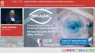
The Eyelib – A multi-ocular robotic platform to automate eye measurements and comprehensive AI-powered diagnosis
- Assouline1,*
1Iena vision, Paris, France
Mikajaki is an emerging startup from Geneva, which designs and manufactures the Eyelib, a multi-ocular robotic platform to automate measurements of eyes, armed with in-house cutting edge AI capabilities for automated eye diagnosis. Mikajaki aims at augmenting services of the ophthalmological clinic by providing a smart and cost-effective platform.
Our solution is based on three main pillars:
- The Eyelib robotic platform: to automate acquisition of objective data, from current prescription through auto-refractometers (pachy, tono, topo, retroillumination, biometry) to OCT for in-depth measurements of the back of the eye
- The Ariane Insight agent: an interactive AI-based chatbot aimed at collecting subjective patient information
- Perform prescreening
- Perform visual test and profile
- Perform initial diagnosis
- An analytics AI-powered reporting platform: A proprietary AI-powered diagnostic platform, providing predictions based on the combination of objective and subjective data. Our AI platform was trained on large sets of data from multiple centers and from specialized cohorts, composed of variable demographics in France (CNIL authorized research, with ~20 partner clinics). All predictions are demonstrable and based on clinical and retrospective studies we performed. We provide predictions such as:
- Subjective refraction
- Risk of glaucoma, angle closure
- Detection and grading cataracts
- Detection of IOL and implant type
- Classification of eye aberrations from retroillumination and OCT
- IOL power predictor
- Eligibility to surgery
To the three pillars above we add an interactive graphical interface, our SmartVision report, which allows the practitioner to review the ensemble of results in an integrative and appealing graphical interface, shortens synthesis of the ensemble of data and facilitates human review of data and diagnosis.
Mikajaki’s unique and integrative approach is distinguished from current technologies by three principal points:
- Automated patient workflow: an integrated platform eliminating the need for patients to move between multiple diagnostic stations
- Multi-modality: combining several optical devices in a single station
- A comprehensive AI-driven fusion of subjective and objective data for diagnosis.
Key features and benefits of Mikajaki’s solution
- Compact Eyelib design, automating the patient journey from registration to data acquisition and finally diagnosis, thus shortening waiting time and sparing resources
- Rapid examination 6-10 minutes per patient
- Collection of objective and subjective data
- High repeatability
- Automated AI-driven diagnosis
- Telemedicine ready
- Interfacing with 3rd party softwares/AI
- Flexible composition of the machines for clinic-tailored needs
- Flexible parameters to adjust to clinical practices
- Ergonomic SmartVision report, adapted to clinic-specific workflows
- Interfacing with Electronic Medical Records
- Centralized internal database
- Motivated and responsive development team
Company website: https://mikajaki.ai/
Supporting documents
-

John Berestka
Northwest Eye, Minneapolis, United States John Berestka
Northwest Eye, Minneapolis, United StatesECS02 – Lightfield Medical
Lightfield Medical: Autonomous digital capture of the entire slit-lamp exam reinvents the heart and soul of ophthalmology
- Berestka, MD1,*
1Cornea, Northwest Eye Clinic, Minneapolis, United States
The ophthalmic slit lamp, the cornerstone of ophthalmology, has remained stagnant for decades. The current slit lamp exam paradigm necessitates the doctor’s physical presence in the room with the patient, making the exam ephemeral and incapable of easy digital capture. Conventional slit-lamp cameras, while useful, slow down the exam, produce inadequate images, and cannot be delegated to technicians. These limitations reduce ophthalmologist productivity and hinder telemedicine in ophthalmology.
For decades, ophthalmologists have delegated retinal and optic nerve imaging to technicians. The missing link in ophthalmology is a solution to automatically capture the entire slit-lamp exam. Lightfield Medical reinvented the slit -lamp from first principles using high speed camera arrays, tunable liquid lenses and a digital light engine. A minimally trained technician simply aligns the device on the pupil and presses a button, allowing the device to take over and capture the anterior segment for the ophthalmologist. The eye doctor can then navigate through the exam in any order using gestures on a phone, tablet or browser. Patients can now easily see their own slit-lamp findings to better understand their own pathology.
As eye care faces a productivity crunch, our medical device will become increasingly essential. The US National Eye Institute predicts a 24% surge in ophthalmology demand by 2023 due to an aging population, yet the ophthalmology supply is expected to decline by 12% during the same period.
Lightfield Medical has developed three generations of prototypes and plans a US product release in late Summer 2025. Our intellectual property portfolio includes four US and one UK patents, with ten additional applications pending. We are led by a team of world-renowned ophthalmologists, including four past presidents of ESCRS, ASCRS, ISRS, and AECOS.
Our device will empower eye doctors to streamline patient care by delegating exam acquisition to technicians. Imagine a scenario where post-operative or brief exam patients are seen by a technician at a satellite office conveniently located near their homes. The exam can be reviewed by an ophthalmologist between surgeries. Findings and instructions can be promptly communicated to the patient via a quick video text message.
Our product will revolutionize eye care by fully documenting the entire slit-lamp exam for the first time. This advancement enables the comparison of eye exams from visit to visit and from doctor to doctor. The standardized lighting and viewing angles will create a homogeneous image database for artificial intelligence.
Company website: www.lightfieldmedical.com
Supporting documents
ESC02 – Lightfield Medical
-

Dr Przemyslaw Korzeniowski
Dr Przemyslaw Korzeniowski
ECS03 – The Next-Gen High-Fidelity Ophthalmic Surgery Simulator
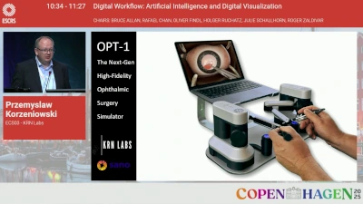
The Next-Gen High-Fidelity Ophthalmic Surgery Simulator
- Korzeniowski1,2,*
1CEO, KRN Labs, 2Medical Imaging and Robotics, Sano Centre for Computational Medicine, Kraków, Poland
KRN Labs is an immersive technology startup specializing in high-fidelity virtual reality (VR) surgical simulators. Founded in 2018 by Dr. Przemysław Korzeniowski, Imperial College London alumni, the company aims to revolutionize surgical training by providing realistic, affordable, and accessible VR solutions for medical professionals worldwide.
Mission and Vision
KRN Labs believes that VR simulation can significantly enhance surgical training by offering a safe, controllable, and configurable environment. This approach eliminates the ethical concerns and high costs associated with traditional training methods involving animals or cadavers. By leveraging VR technology, KRN Labs strives to improve the educational experience without putting patients at risk.
Product Portfolio
KRN Labs has developed a suite of VR surgical simulators tailored to various training needs:
SurgicaVR: Designed for robotic and laparoscopic surgery training, SurgicaVR utilizes consumer-grade VR headsets like Meta Quest 2/3 and Apple Vision Pro. It currently features a comprehensive cholecystectomy procedure, with plans to expand its library with diverse surgical scenarios and real-time feedback systems.
SurgicaVR Pro: Building on SurgicaVR, this simulator offers ultra-realistic graphics and detailed procedural scenarios tailored for institutions and individual practitioners. It includes scalable training modules and performance tracking tools, making high-fidelity laparoscopic training more accessible.
SurgicaVR ProHaptics: This advanced simulator integrates Follou’s Haptic Avatar technology, providing precise force feedback for an authentic surgical experience. It allows medical professionals to train in laparoscopic procedures with unparalleled realism.
We also developed a cataract simulator for the leading global eye-care company.
Technological Innovation
KRN Labs addresses the limitations of conventional surgical training by offering VR simulators that replicate real-life surgical conditions. These simulators provide a safe and ethical training environment, enabling clinicians to practice repetitively without risking patient safety. The integration of haptic feedback technology further enhances the realism of the training experience.
KRN Labs continues to push the boundaries of medical training by developing innovative VR solutions that enhance the skills of healthcare professionals, ultimately aiming to improve patient care outcome.
Company website: https://krn-labs.com
Supporting documents
-

Enrique Vega
Enrique Vega
ECS04 – The Eye Reimagined
The Eye Reimagined: A Lens-Embedded Platform for Real-Time Care and Beyond.
Delivering real-time visual correction, health monitoring, and digital interaction through adaptive optics, embedded electronics, and seamless connectivity.
- Vega1,*
1Azalea Vision, Ghent, Belgium
Azalea Vision is developing the first medical-grade smart lens platform that adapts, connects, and delivers real-time clinical value. Spun off from imec and Ghent University, Azalea integrates adaptive optics, liquid crystal technology, and embedded electronics within a scleral lens to treat complex vision conditions like keratoconus and presbyopia. Backed by leading investors and supported by public innovation grants, Azalea’s technology dynamically corrects vision and communicates with smartphones or wearables via NFC. Future applications include tear-based biosensing, precision drug delivery, and integration into AR/VR systems—redefining vision as an intelligent, connected health interface.
Extended Summary:
Azalea Vision is pioneering the future of connected ocular health through the development of the first medical-grade, intelligent lens-embedded system designed for real-time clinical impact.
Founded in 2021 as a spin-off from imec and Ghent University, Azalea combines deep expertise in microelectronics, biomedical optics, and medical device innovation. The company is backed by leading HealthTech, MedTech, and DeepTech investors—including imec.xpand, Elaia Partners, Sensinnovat, and SPRIM SGI—and supported by public innovation grants such as the European Innovation Council (EIC) and VLAIO.
At the core of Azalea’s platform is a breakthrough smart lens technology that integrates adaptive optics, liquid crystal filters, custom ASICs, and NFC-enabled communication—all engineered within a scleral lens format. This technology mimics the natural responsiveness of the human eye, adapting in milliseconds to changes in light, focus, and motion, while remaining seamlessly connected to external devices like smartphones or smartwatches.
Azalea’s first clinical application targets patients with corneal irregularities, such as keratoconus and irregular astigmatism, conditions often underserved by existing static or surgical solutions. The lens provides dynamic, personalised correction of higher-order aberrations (HOAs), improving visual acuity and quality of life for patients who cannot benefit from standard interventions.
And Azalea’s platform goes far beyond correction. It is built to scale across a pipeline of future applications, including:
– Tear-based biosensingfor conditions like glaucoma, dry eye, and diabetes (via glucose, IOP, etc.)
– Precision drug deliverydirectly through the lens
– Adaptive optics for AR/VR integrationand real-time visual enhancement
By reimagining the eye as a connected interface—not just a sensory organ—Azalea Vision is shaping a future where vision is intelligent, personalised, and deeply integrated into digital health ecosystems.
We’re not just building a lens.
We’re building the next platform in ocular care.Company website: www.azaleavision.com
Supporting documents
-

Almog Aley-Raz
Almog Aley-Raz
ECS05 – CorNeat Vision
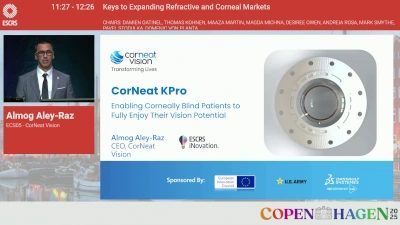
CorNeat Vision: How a novel synthetic biomaterial can impact global blindness – latest clinical updates from the EverPatch+ launch, CorNeat KPro clinical trial, and first implantations of the eShunt.
- Aley-Raz1,*
1R&D, CorNeat Vision Ltd, Raanana, Israel
CorNeat Vision was established 9 years ago with the primary goal of developing and commercializing the CorNeat KPro, an artificial cornea. This implant and its novel implantation procedure enable corneally blind patients to completely and immediately rehabilitate their vision following a relatively straightforward procedure without the need for donor tissue, which is limited in supply. The CorNeat KPro’s long-term goal is to significantly improve the current standard of care (keratoplasty) by providing a scalable solution with predictable and long-lasting optical performance equivalent to a perfect cornea.
The underlying material technology developed by the company, EverMatrix, enables the permanent integration of a synthetic lens with resident ocular tissue and presents a significant opportunity. EverMatrix is the first non-degradable biomaterial that seamlessly and permanently embeds with living tissue without triggering a significant chronic inflammatory response. Its durability and flexibility, combined with its biomechanical integration capabilities, support a variety of use cases with applications spanning every field of surgery.
CorNeat Vision is at the forefront of ophthalmic innovation with its flagship product, the CorNeat KPro, which is progressing in the clinical phase with promising and impactful results. The company also offers a unique synthetic permanent scleral reinforcement matrix for ocular surface surgeries, EverPatch+ (FDA cleared). The device is being soft-launched to the market, gradually replacing tissue patches used in glaucoma shunt and other ocular surface surgeries. The company’s third product, eShunt, is a unique glaucoma drainage device that extends minimally invasive surgeries to severe and refractory glaucoma patients. The device leverages the EverMatrix technology to integrate with the eyewall and avoid encapsulation around its outlet, which is uniquely positioned in the orbital fat. The eShunt is currently in first-in-human implantations. Initial results obtained in a clinical trial in glaucomatous dogs demonstrate the device’s lasting capability to reduce IOP to the low teens.
Company website: www.corneat.com
Supporting documents
-

William Link
William Link
ECS06 – EyeDura Therapeutics
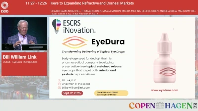
EyeDura Therapeutics – Transforming Anterior and Posterior Ocular Drug Delivery
- Link1,*
1EyeDura Therapeutics, New York, United States
EyeDura Therapeutics is an early-stage ophthalmic pharmaceutical company developing preservative-free topical sustained release eye drops that target both anterior and posterior eye conditions. Current approaches to ocular drug delivery continue to face critical challenges due to various barriers in the eye. For anterior conditions, patients endure frequent dosing regimens that create peaks and troughs in drug concentration. For posterior conditions, patients must undergo invasive injections or implants. These approaches lead to poor patient compliance, significant logistical burdens, and high costs to the payors.
EyeDura is poised to transform how topical eye drops are used. A single drop of the topical sustained release (TSR) formulation will deliver therapeutic levels of drug for seven days or longer. The TSR vehicle is compatible with most drugs currently used in ophthalmic products including both small molecules and biologics.
EyeDura formulation has a novel mechanism of action. During a blink, the aqueous layer is selectively removed while the lipid layer stays on. The formulation resides on the lipid layer and therefore remains on the surface for extended periods and provides a stable depot. The product is preservative free.
Our lead asset is Topical Sustained Release formulation of Insulin and it targets neurotrophic keratitis (NK), with an IND filing planned for 1Q2026.
We ran a probing trial in Central America where we treated 10 patients with persistent epithelial defects with the topical sustained release insulin formulation. After two drops per week of treatment with our formulation, the epithelial defects healed completely in all ten cases within a month. This is a remarkable finding and needs further clinical studies in NK patients.
We’ve also demonstrated our platform’s ability to deliver medication to posterior eye structures. We dosed five patients with the topical sustained release insulin drops at two drops per week and were able to achieve therapeutic levels of insulin up to 5 ng/mL in the vitreous. Literature shows that insulin restores retinal ganglion cell functional connectivity and promotes visual recovery in glaucoma.
We have a dual path for revenue generation. The first is to develop our internal assets beginning with NK and explore licensing opportunities for the asset. In parallel, we plan to develop strategic collaborations with ophthalmic companies to incorporate their drug into our platform technology.
Our technology is protected by strong global IP with eight issued and eight published patents extending through 2044. Our board, leadership team, and advisors bring deep expertise in ophthalmology, drug development, and commercialization.
We are seeking a $25M Series A investment and a lead investor to advance our NK program through clinical development, Build our development, regulatory, and operations capabilities, and finally, expand our strategic partnerships.
Company website: www.eyedura.com
Supporting documents
-

Rania Maklad
OCUWELL Rania Maklad
OCUWELLECS07 – OCUWELL
OCUWELL: Transforming Ocular Diagnostics with Precision
- A. Maklad1,*
1Ocuwell Limited, Liverpool, United Kingdom
The Problem: Problem
Corneal disorders, including high myopia, astigmatism, cataract, keratoconus, dry eyes, and corneal dystrophies affect over 850m people worldwide. These conditions contribute to an estimated 43 million cases of blindness and 217 million cases of moderate to severe visual impairment.
Diagnosis and management of corneal disorders necessitates access to corneal topography devices. However, these devices are often expensive and bulky, unsuitable for children, bedridden and psychiatric patients, and require travel to specialised clinics, resulting in delayed treatment. Our 2021 survey showed that over 80% of UK optometry clinics and 40% of specialised tertiary clinics lack access to these devices – and the situation is worse in developing countries, leading to 28% of patients with corneal disorders remaining undiagnosed in the early stages. Furthermore, over 60% of newly diagnosed cases in some developing countries are classified as severe upon initial presentation, resulting in serious permanent visual acuity losses.
Affordable, portable corneal topography devices are urgently needed for the optimal management of corneal abnormalities, including early detection of ectasia, optimising soft and rigid contact lens fitting (myopia control), and follow-up surgical interventions.
OCUWELL’s innovative technology aims to address this global health need.
Importance
Timely diagnosis and effective management of eye conditions can significantly improve the quality of life for millions of people, prevent vision loss, and reduce the burden on healthcare systems. The Lancet Global Health Commission published their comprehensive findings in 2021[9], illustrating the extent of the burden and the health inequalities present. By providing an affordable and accessible solution, OCUWELL can empower healthcare providers to make informed decisions and deliver personalised care to patients, filling the current gap in the market and meeting the global demand for advanced eyecare solutions.
The Solution: Our technology aims to significantly reduce the risk of blindness and visual impairment caused by corneal disorders such as high myopia, astigmatism, keratoconus, and corneal dystrophies, with future development plans including the detection of dry eye disease. By equipping healthcare professionals with advanced, accessible diagnostic tools, we are working to transform the standard of care for corneal conditions, address healthcare inequalities, and improve access to timely, remote diagnostics and progression monitoring — directly supporting UN Sustainable Development Goal 3: Good Health and Well-Being.
Our unique selling proposition lies in delivering full corneal topography through a low-cost, portable, hand-held device that can be used in any setting. This enables early detection and management of conditions that represent a £1.25 billion global market opportunity.
By replacing bulky hardware components with sophisticated software modules, our device offers highly precise analysis at just 60% of the cost of the most affordable systems currently on the market — making high-quality eye diagnostics more accessible than ever.
Company website: https://www.ocuwell.com/
Supporting documents
-

Susana Marcos
Susana Marcos
ECS08 – Simulation of multifocal corrections in the clinic
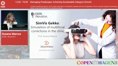
Simulation of multifocal corrections in the clinic
- Marcos1,2,*
1University of Rochester, Rochester, United States, 2CSIC, Madrid, Spain
PROBLEM & UNMET NEED
Presbyopia is a non-preventable, gradual, and irreversible eye condition characterized by the loss of the ability to focus on near objects. It causes significant visual symptoms in over 80% of the population above the age of 50—more than 2 billion people by 2024. Similarly, cataracts are age-related opacifications of the eye’s natural lens. As of 2025, they affect
over 50% of individuals over 75 years old—approximately 110 million people. Cataract surgery is among the most frequently performed procedures worldwide, with around 30 million surgeries conducted annually (second only to childbirth).
Surgery is the standard treatment for both conditions. It involves replacing the eye’s crystalline lens with an artificial intraocular lens (IOL). There are over 100 different IOL models available from more than 30 manufacturers, each offering a distinct visual experience. However, the effect of each lens is difficult to convey, and patients cannot preview their
postoperative vision. As a result, lens selection is typically made through a simple conversation between doctor and patient, without incorporating the patient’s feedback or preferences.
This suboptimal selection process leads to reduced postoperative satisfaction and poses a significant barrier to the adoption of multifocal lens corrections.OUR TECHNOLOGY & SOLUTION
SimVis technology is based on temporal multiplexing using tunable lenses (TLs), which change optical power in response to electric signals. 2EyesVision’s proprietary technology allows presbyopic and cataract patients to experience the real world through simulations of existing intraocular lenses—enabling them to “try on” different visual corrections prior to
surgery and provide feedback that informs the selection of the most appropriate IOL.
Our product, SimVis Gekko2, is a visual simulator for presbyopia and cataract surgery that, for the first time, enables patients to test multifocal corrections before implantation. It is a binocular, head-mounted, see-through optical device that provides a natural visual experience.Company website: www.2eyesvision.com
Supporting documents
-
Milind Pande
Milind Pande
ECS09 – CustomLens Ai

CustomLens Ai: Personalising Vision, Scaling Precision
- Pande1,*
1Ophthalmology, Vision Surgery & Research Centre, North Ferriby , United Kingdom
CustomLens.ai is a cloud-based comprehensive lens surgery expert system designed to automatically generate complete surgical prescriptions inclusing intraocular lens (IOL) selection and surgical planning through explainable AI. CustomLens Ai is proud to be part of Microsoft for Startups.
Built by surgeons, for surgeons, it delivers personalised surgical plans and multidimensional functional vision outcomes tailored to each patient’s unique biometric profile and the operating surgeon’s preferences. It is a full lens surgery system which provides full outciomes analysis, alternative outcoime scenarios. It aims to be the leading AI company in lens surgery by continuing to develop innovations in AI which make an actual difference to unaided vision outcomes.
At the heart of CustomLens.ai is a proprietary, symbiolic AI based expert system that replicates expert-level decision-making. It integrates historical outcomes, preoperative diagnostics, and nuanced surgical judgement into a single intelligent interface—bridging the gap between standard formulae and real-world surgical practice. It also provides for multidimensional functional vision outcomes presented in a patient friendly education system called VisionCoach AI.
CustomLens Ai represents the next step in surgical intelligence—where data, human judgement, and intelligent automation come together to deliver better vision, at scale. It makes acheiving premium vision outcomes easy for every surgeon.
Company website: https://customlens.ai
Supporting documents
-

Olivier Benoit
Olivier Benoit
ECS10 – Glaucoma Surgery Reinvented
Glaucoma Surgery Reinvented: Ab Externo Supraciliary Approach with No Bleb, No Cleft, and Preserved Anterior Chamber
- Benoit1,*
1CILIATECH, Annecy, France
A Breakthrough Approach to Glaucoma Treatment: “No Anterior Chamber, No Bleb, No Cleft”
CILIATECH has developed a truly novel ab externo surgical method for treating glaucoma, centered around a next-generation implant called INTERCIL, recently CE-marked for both primary open-angle glaucoma (POAG) and primary angle-closure glaucoma (PACG).
Unlike existing techniques—including filtering surgeries and angle-based minimally invasive glaucoma surgeries (MIGS)—this new ab externo approach does not involve the anterior chamber, creates no filtration bleb, and requires no angle disruption. INTERCIL is uniquely postionned in the supraciliary space, engaging the uveoscleral outflow pathway while preserving the eye’s natural anatomy. This innovative positioning enables strong and durable intraocular pressure (IOP) reduction while minimizing postoperative risks.
INTERCIL acts as a uveal spacer, creating a controlled separation in the supraciliary space to promote aqueous outflow via both anterior and posterior uveoscleral pathways.
Clinical results from a pilot study involving 56 patients (29 POAG, 28 PACG) are highly promising. Using the final device design and updated surgical technique:
- Mean baseline IOP: 23.5 ± 1.7 mmHg and 1.8 ± 0.8 medications
- Mean IOP at 12 months (n=44): 14.1 ± 3.0 mmHg and 0.2 ± 0.6 medications, 86% of patients are medication free
- Mean IOP at 24 months (n=39): 13.9 ± 3.1 mmHg and 0.3 ± 0.7 medications, 77% of patients are medication free
- No difference in efficacy between POAG and PACG groups
The procedure showed an excellent safety profile:
- No serious adverse events
- No cases of clinical hypotony, inflammation, implant migration, or foreign body reaction
- Smooth surgical learning curve with accurate implant placement
Despite no bleb, no cleft, and no anterior chamber intervention, INTERCIL delivers IOP reduction in the low mid-teens range with a great safety profile. This ab externo cilioscleral interposition technique offers a powerful, safe alternative for patients who need strong IOP reduction but wish to avoid the risks of invasive filtering procedures.
Company website: www.cilia.tech
Supporting documents
-

Shawn O’Neil
Shawn O’Neil
ECS11 – FLIGHT by ViaLase
FLIGHT by ViaLase: The First Noninvasive Femtosecond Laser Procedure for Glaucoma
- England1,*
1ViaLase, Inc, Aliso Viejo, United States
ViaLase is a clinical-stage medical device company pioneering FLIGHT (Femtosecond Laser Image-Guided High-Precision Trabeculotomy), the first and only noninvasive femtosecond laser procedure designed to treat primary open-angle glaucoma. FLIGHT precisely creates a channel through the trabecular meshwork to enhance aqueous outflow—without requiring incisions, implants, or entry into the anterior chamber.
ViaLase was founded by Dr. Tibor Juhasz, a physicist, inventor, and entrepreneur widely recognized for introducing femtosecond laser technology to ophthalmology. He was a founding figure behind IntraLase (bladeless LASIK) and LenSx (laser cataract surgery), both of which transformed standards of care and were acquired by major strategics. With ViaLase, Dr. Juhasz brings his track record of category-defining innovation to the field of glaucoma.
The ViaLase system integrates high-resolution OCT imaging with femtosecond laser precision, enabling image-guided, nonincisional targeting of outflow structures. The procedure is designed to be performed outside the operating room and supports repeatability if additional IOP reduction is needed.
ViaLase is addressing a critical unmet need between medical therapy and surgery, offering a novel, nonincisional solution for millions of patients with mild to moderate glaucoma.
Company website: www.vialase.com
Supporting documents
-

Henrik Nagel
Henrik Nagel
ECS12 – MistGo®
MistGo® is an innovative alternative to the conventional eye drop bottle, designed to help patients administer their eye medication in a more precise and user-friendly way. A better user experience translates into higher patient compliance, healthier eyes, and higher quality of life for patients.
- Nagel1,*
1Denmark Technical University, Copenhagen, Denmark
The Problem: EYE-GO addresses a large unrealized potential in pharmaceutical companies – and at the same time, we contribute to patients’ quality of life!
Currently, eye patients have to put up with the familiar eye drop bottles. Data shows that up to 75% do not receive the right treatment with the eye drop bottle for various reasons. Among other things, because it is too much of a hassle with the eye drop bottles. As a result, patients probably do not achieve satisfactory health results from the treatment.
However, it is also a huge problem for the pharmaceutical company that the desired treatment effect is not achieved, and business potential is missed, ultimately for society, which ends up with much higher costs for the healthcare system than necessary.
The Solution: EYE-GO’s solution is a new generation of ophthalmic drug delivery systems.
EYE-GO’s solution removes all the barriers known from eye drop bottles and realizes the full potential of the drug, so the actual result is the same as what everyone expected.Our solution is superior in the following parameters to conventional eye-drop bottles:
– Micro-dosing with precision on the cornea, 6 µL, no overmedication. Eye-drop bottles, and vial dose between 30-50 µL.
– Ease of use and comfort for the patient, with the head in any position.
– Independent of the patient’s motor abilities. All other delivery systems depend on the patient’s ability to hold the dosing unit in the correct position in front of the eye/head.
– The risk of cross-contamination is very low.
– Preservative-free drug compatibility.
– Viscosity window, 1 – +500 cP. Other delivery systems that offer preservative-free drug compatibility have a narrow window of 1 – 15 cP.
Supporting documents
2024
-

Ran Yam
NovaSight, Airport City, Israel Ran Yam
NovaSight, Airport City, IsraelECS01
NOVASIGHT IS A GROWING MEDICAL DEVICE COMPANY WITH ITS FLAGSHIP PRODUCT – CURESIGHT™ – FDA CLEARED AND CE MARKED REMOTE DIGITAL EYE TRACKING BASED AMBLYOPIA TREATMENT SYSTEM, THE ONLY DIGITAL DICHOPTIC THERAPY FOR AMBLYOPIA PROVEN TO BE NON-INFERIOR TO THE PATCH.
R.Yam1,*
1NovaSight, Airport City, Israel
Established in 2016, NovaSight is a growing ophthalmology medical device company with a vision to change the lives of millions by introducing eye-tracking-based vision care solutions and data analytics into fun and engaging medical devices with an emphasis on the unique needs and attention spans of children. NovaSight is focused on three main segments: Amblyopia treatment, Myopia prevention and Vision diagnostics.
Commercially available in the US and Italy since early 2023, CureSight™ is an FDA-cleared and CE-marked amblyopia treatment system, designed to replace the traditional eye patching. The treatment is carried out while the patient watches their favorite streamed content from the comfort of their home. To date, CureSight is the only solution that has proven effective in a pivotal study against the gold standard patching in children.
By tracking the gaze position of the dominant eye in real-time, the CureSight system blurs its center of vision, while keeping the rest of the image sharp, providing the amblyopic eye with a normal, high-contrast, image. This stimulates the brain to complete the blurred image from the amblyopic eye, improving its acuity and developing stereoacuity as the eyes learn to work together. In addition, the CureSight system is connected to a dedicated cloud platform which allows the physician to monitor treatment progress and compliance remotely.
Since launch earlier last year, CureSight had gained strong adoption by both eye care providers and patients, with over 250 eye care providers already enrolled in the CureSight Referral Program. NovaSight is continuously onboarding additional prominent clinics, resulting in the active availability of the CureSight Referral Program in 35 states with over 650 patients referred so far.
The company’s goals include a global launch of CureSight, Europe launch of the EyeSwift PRO, and further development of additional products.
Company website: https://nova-sight.com/
Supporting documents

-

Dimitri Chernyak
Intelon Optics, Woburn, United States Dimitri Chernyak
Intelon Optics, Woburn, United StatesECS02
MEASURING OCULAR BIOMECHANICS BY BRILLOUIN IMAGING
D.Chernyak1,1,*
1Intelon Optics, Woburn, United States
Intelon Optics is a medical device company focusing on the development and commercialization of the Brillouin Optical Scanner System (BOSS™), a technology that enables non-invasive, next-generation point-by-point biomechanical imaging of living tissue and cell structures, with initial applications in Eye care (Ophthalmology) and Fertility (IVF – In Vitro Fertilization).
In Ophthalmology, BOSS delivers novel diagnostic solutions to eye care professionals for high-level decision-making confidence based on biomechanical properties of the eye. Primary clinical applications are early detection of corneal deseases, refractive surgery screening and customization, reduction of post-cataract surgery astigmatism with custom AKs and LRIs. BOSS core technology, the ultra-narrow bandwidth laser and the Brillouin spectrometer, provides a common platform engine for all its medical applications.
Company website: www.intelon.com
Supporting documents
-

John C. Berestka
Northwest Eye, Minneapolis, United States John C. Berestka
Northwest Eye, Minneapolis, United StatesECS03
LIGHTFIELD MEDICAL: AUTOMATED CAPTURE OF THE SLIT LAMP EXAM—REINVENTING AN 85- YEAR OLD TECHNOLOGY
J. C. Berestka1,*
1NORTHWEST EYE, MINNEAPOLIS, United States
The ophthalmic slit lamp–the heart and soul of ophthalmology–has not seen any innovation in decades. The current slit lamp exam paradigm requires the doctor to be present in the room with the patient. The slit-lamp exam is ephemeral and cannot be digitally saved. Existing slit lamp cameras slow down the encounter, provide inadequate images and cannot be delegated to a typical technician. Limitations in current slit-lamp technology preclude adequate telemedicine in ophthalmology and throttles ophthalmologist productivity.
Ophthalmologists have been able to delegate retinal and optic nerve imaging to technicians for decades. The missing link in ophthalmology is a way to automatically capture the slit-lamp exam. Our medical device uses proprietary technology to capture the entire slit-lamp exam. A minimally trained technician only needs to align the device on the pupil and press a button. The device takes over and then images the anterior segment for the ophthalmologist. The eye doctor can then navigate through the the exam in any order using gestures on an iPhone and iPad. Patients can now see their own slit-lamp findings either in the office or in the comfort of their home.
Our patented technology (4 US and 1 UK patent granted) will allow eye doctors to more efficiently see patients by delegating exam acquisition to technicians. imagine a scenario where postop or brief exam patients are seen by a technician at a satellite office close to their home. The exam could later be reviewed by an ophthalmologist in between surgeries. Findings and instructions could be communicated back to the patient by a quick video text.
Our medical device will be increasingly needed as eye care is about to experience a productivity crunch. The US National Eye Institute predicts a 24% increase in ophthalmology demand by 2023 due to an aging population, yet the ophthalmology supply is expected to decline by 12% during the same period.
Our product will allow the entire slit-lamp exam to be fully documented for the first time. With our technology eye exams can now be compared from visit to visit, and from doctor to doctor. The standardized lighting and viewing angles will create a homogenous image database for artificial intelligence.
Patients can be given a copy of their eye exam to view on their phones, in much the same way that patients can now scroll through their MRI exams in apps such as MyChart in the US.
Currently, less than 1% of eye exams are done through telemedicine, because until now there was no good way to capture the examination of the front of the eye.
Our device will cost half as much as a wide-field retina camera such as Optos, providing value to patients and doctors. Our medical device could bring eye care to remote areas of the world that are currently underserved by eye doctors.
Company website: https://lightfieldmedical.com
Supporting documents
-

Phil Norris
machineMD, Bern, Switzerland Phil Norris
machineMD, Bern, SwitzerlandECS04
MACHINEMD LAUNCHES NEOS™ – THE WORLD’S FIRST NEUROPHTHALMOSCOPE
P. Norris1,*
1machineMD, Bern, Switzerland
machineMD is a medical device startup innovating at the intersection of neuroscience and ophthalmology. The company was founded in 2019 as a spin-off from the Eye Movement Laboratory of the University Hospital of Bern, Switzerland, where it retains headquarters, and now also has a subsidiary in Boston, USA.
We believe objective eye exams increase healthcare provider efficiency, reduce clinical error and improve patients’ quality of life.
Launched in 2024, machineMD’s flagship device is neos™ – the world’s first neurophthalmoscope – an automated medical device that supersedes the need for manual assessments of eye movements and pupillary function that are conducted daily by optometrists, comprehensive ophthalmologists, and general neurologists.
Current starndard of care is manual, time-intensive, subjective examinations. Misdiagnosis in primary and secondary care is evident in 50% of neuro-ophthalmic referrals, leading to significant harm for 26% and unnecessary procedures for 80% of those patients. Median referral time to a neuro-ophthalmologist is 200 days, with a recognized shortage in filling these subspecialty fellowships and full-time positions.
To address this unmet need, neos™ enables medical assistants to perform a fast, objective, standardized examination of eye movements and pupillary function, leveraging an industry-leading VR headset with gamified stimuli. High-frequency eye tracking enables neos™ to empower primary, secondary, and tertiary care providers with quantitative reports compatible with electronic health record systems.
Abnormalities in eye movements and pupillary function are closely linked to neurological disorders, with the potential to serve as important digital biomarkers for the diagnosis and monitoring of inflammatory and neurodegenative disorders. Together with neurologists, machineMD’s solution is already used to study people living with Multiple Sclerosis, Parkinson’s Disease, Myasthenia Gravis, and beyond.
Company website: www.machinemd.com
Supporting documents
-

Nir Israeli
EyeMed Technologies Ltd., Ramat Gan, Israel Nir Israeli
EyeMed Technologies Ltd., Ramat Gan, IsraelECS05
VALENS – POST OPERATIVE ADJUSTABLE INTRAOCULAR LENS
N. Israeli1,*
1EyeMed Technologies Ltd., Ramat Gan, Israel
EyeMed Technologies Ltd. is a medical device start-up company that focus on the Cataract surgery market, specifically, on the Premium IOL segment. The company develops an intra-ocular lens, called VaLens™, which enables the physician to precisely adjust the power of the lens in a non-invasive postoperative, office-based, simple treatment. EyeMed’s vision is to guarantee Emmetropia (best vision possible) and “glasses off” for every lens implanted patient. There is a significant number of cases in which, post-Cataract surgery, the patient is left with a refractive error causing the patient a deficiency in his eyesight because of misalignment of the focal point of the lens. Today there are solutions for residual refractive errors due to IOL misposition, however they are either a burden on the patient (whether it is to wear eyeglasses or to settle for a deficient vision) or they require additional surgical procedures with all attached risks and resources. The VaLens™ innovative solution is intended to be implanted in every Cataract surgery embedding a premium lens and to enable simple solution to all those patients that need a postoperative adjustment after implantation.
Cataract surgery is the most frequent surgical procedure in medicine today, with about 30M surgeries performed annually. The premium IOL segment sales projection, in 2024, is a $3.1B annual revenues. Studies show that 28.8% of Cataract implanted patients have a residual refractive error bigger than 0.5D and 7% more than 1.0D, errors that should be corrected. Ophthalmologists have been implanting multifocal and premium technologies since decades, and still this technology has been used in only 3-9% of lens implantations world-wide. Percentage of usage is not growing even there are lots of efforts in this direction, mostly because surgeons do not feel confident enough to be able to guarantee their valuable patients that they will have emmetropia post-surgery. Additionally, surgeons prefer not to deal with the likelihood of post-surgery touchups that require additional time, resources and are a burden from a patient perspective. EyeMed’s device, the VaLens™, will deliver surgeons the confidence they need with reduced resources and ensure patients with the best results.
The VaLens™ foldable adjustable IOL consists of a nitinol mechanism placed in a haptic-optic cradle. Heating actuators on the nitinol mechanism with a continuous green laser achieve controlled movement of the mechanism and optic. Activation occurs in controlled steps: rotation in 1-degree steps and anterior posterior movement in 0.25D steps with a range of +1.50D. The IOL was tested in vitro and in vivo in a rabbit eye. Foldability and unfolding were demonstrated through a 2.6-mm cartridge.
Company website: www.eyemed.co.il
Supporting documents
-

Mikhail V. Tsurkan
TissueGUARD GmbH, Dresden, Germany Mikhail V. Tsurkan
TissueGUARD GmbH, Dresden, GermanyECS06
SUBSTITUTION REGENERATIVE THERAPIES FOR LIMBAL, ENDOTHELIAL, AND RETINAL DYSFUNCTION
M. V. Tsurkan1,*
1TissueGUARD GmbH, Dresden, Germany
TissueGUARD GmbH is a privately-funded biotechnology company dedicated to developing and producing advanced hydrogel materials as medical devices. Our mission is to create innovative biotechnology solutions for tissue and organoid engineering as regenerative therapeutics.
At TissueGUARD, we have developed proprietary hydrogel materials that are fully defined, sacrificially degradable, and capable of supporting the growth of almost any cell type. Our unique technology allows for the on-demand removal of the device without harming the formed organoids or tissues, enabling the formation of physiological-sized, donor-like grafts. This scaffold-free approach simplifies tissue and organoid implantation, enhances therapeutic outcomes, prevents implantation-associated complications, and significantly reduces regulatory certification burdens in both the EU and the USA.
Our cutting-edge bioengineering capabilities have led to remarkable advancements in ocular regenerative approaches. Our technique enables the formation of corneal endothelium grafts with preformed Descemet-like membranes. It has also been shown to form retinal pigment epithelium (RPE) with preformed Bruch’s membrane. These pioneering tissue graft technologies have demonstrated potential in stabilizing surgical implantation and improving outcomes in ophthalmologic procedures. TissueGUARD’s technology not only addresses the critical shortage of donor tissues but also paves the way for comprehensive regenerative therapies for limbal, endothelial, and retinal dysfunctions.
Company website: https://tissueguard.de/
Supporting documents
-

Greg Kunst
Aurion Biotech, Seattle, United States Greg Kunst
Aurion Biotech, Seattle, United StatesECS07
TRANSFORMATIONAL CELL THERAPY FOR CORNEAL ENDOTHELIAL DISEASE
G. Kunst1,*
1Aurion Biotech, Seattle, United States
Aurion Biotech is a clinical-stage biotech company, whose mission is to restore vision to millions of patients with life-changing regenerative therapies. It received the prestigious Prix Galien award for best start-up in biotech. Its first candidate, AURN001, is for the treatment of corneal edema secondary to corneal endothelial disease, and the first clinically validated cell therapy for corneal care, having received regulatory approval in Japan. The Company has completed enrollment and dosing of subjects in its Phase 1/2 clinical trial in the U.S. and Canada, and expects to share topline data in early 2025. Privately held, Aurion Biotech is backed by Deerfield, Alcon, Petrichor, Flying L Partners, Falcon Vision / KKR, and Visionary Ventures.
Company website: www.aurionbiotech.com
Supporting documents
-

Sunil Shah
Aston University, Birmingham, United Sunil Shah
Aston University, Birmingham, UnitedECS08
A NOVEL HAND HELD UVC EMITTING DEVICE FOR TREATING INFECTIOUS KERATITIS CAUSED BY A WIDE RANGE OF MICROBES THAT IS SIMPLE AND RAPID TO USE IN CLINIC WITHOUT ANY RISK OF INCREASING RESISTANCE THROUGH SPARING THE USE OF TOPICAL ANTIMICROBIAL AGENTS
S. Shah1,*
1Aston University, Birmingham, United Kingdom
We are developing a patented ultraviolet C device to kill corneal infections in humans and animals. The device can be hand held and slitlamp mounted. The infected corneal ulcer is treated for 5 seconds. UVC is effective against bacteria, viruses and fungi and hence can be used for any type of infection either alone or in addition to conventional treatment.
Technology: UVC is well known to be antimicrobial and has been used for sterilisation in many industries. More recently LEDs have become available of the appropriate wavelengths to enable a hand held device to be made. Extensive work has shown that with the dose of UVC being used, there is a 100% kill rate in animal studies with no lasting damage to healthy tissue. There is no microbial resistance.
Patient pool: 600,000 human corneal infections and 6 million animals (primarily cats and dogs) corneal infections per year in USA and Europe. In countries like India, 2 million monocular blindness from corneal infections.
IP Protection: First layer patents granted and second layer pending.
As regulatory hurdles are less with the animal market, plan is to start in that space initially while the human studies and regulatory process is on going. Animal income to help fund human studies.
Device time line: Prototype available Q3 2024 and commercial device available Q1 2025.
Team: A combination of practicing ophthalmologists, academic researchers, ophthalmology entrepreneur, veterinary surgeons and accountant/entrepreneur in multiple spaces
USP: Novel, immediate effect and cost effective in a market that has not had significant changes for many years.
The aim of the presentation would be to help raise awareness and to meet potential investors or partners both for distribution and/or acquisition.
Company website: www.photon-therapeutics.com
Supporting documents
-

Marco Lombardo
Studio Italiano di Oftalmologia, Rome, Italy Marco Lombardo
Studio Italiano di Oftalmologia, Rome, ItalyECS09
UNVEILING INCISION-FREE THERANOSTIC TECHNOLOGY FOR THE CORRECTION OF VISUAL DISORDERS
M. Lombardo1,*
1Studio Italiano di Oftalmologia, Rome, Italy
Regensight is pioneering a groundbreaking advancement in eye care with theranostics, a cutting-edge method that revolutionizes the treatment of visual disorders using an incision-free approach. Theranostic-guided UV-A light photo-reshaping leverages the principles of theranostics and the therapeutic potential of UV-A light to non-invasively reshape corneal tissue. This promising technique can treat a variety of visual disorders across all age groups. Theranostics combines diagnostic and therapeutic functions into a single sophisticated platform, involving advanced molecular imaging and targeted UV-A light photo-reshaping process delivered simultaneously. The key aspects of how this technology can revolutionize ophthalmic care include:
- Targeted Drug Delivery: theranostics enables fast and precise targeted delivery of riboflavin to specific regions of the corneas, ensuring optimal therapeutic concentration and enhancing treatment efficacy under UV-A light mediated photo-reshaping procedures.
- Real-Time Monitoring: the integration of diagnostic and therapeutic functions allows for real-time monitoring of treatment efficacy. Molecular imaging technologies provide immediate feedback on how the cornea is responding to treatment, enabling adjustments to be made quickly and accurately.
- Minimizing Invasive Procedures: by eliminating the need for surgical incisions, theranostics reduces the risk of complications, such as infections and corneal scarring.
- Enhanced Patient Experience: patients undergoing incision-free theranostic procedures experience less discomfort and shorter recovery times compared to traditional surgical methods. The non-invasive nature of these procedures can reduce patient anxiety, improve overall satisfaction with care and increase demand for refractive treatments.
The technology has undergone pre-clinical and clinical validation demonstrating the theranostic platform’s capability to predictively personalize therapeutic outcomes for keratoconus. The multicenter, randomized, masked, ARGO trial (NCT05457647) validated the capability of the theranostic platform to treat keratoconus and predict clinical outcomes with 91% accuracy and 95% precision. The clinical trial outcome are published in Ophthalmology, the top-ranked journal in the field. This evidence marks a significant advancement towards predictive personalized care for keratoconus, delivering precise and effective results that will benefit patients and society.
Regensight is now advancing with an innovative platform harnessing the power of theranostics to correct the most prevalent visual disorders across all age groups. The new platform aims to improve uncorrected visual acuity without the need for invasive surgery, particularly in individuals with low-grade myopia and presbyopia. Regensight is seeking investment from corporate investors to support the clinical development of this new platform.
Company website: www.regensight.com
Supporting documents
-

Susana Marcos
CSIC, Madrid, Spain; University of Rochester, Rochester, United States Susana Marcos
CSIC, Madrid, Spain; University of Rochester, Rochester, United StatesECS10
VISUAL SIMULATION TECHNOLOGY FOR MULTIFOCAL CORRECTIONS BEFORE CATARACT OR PRESBYOPIA CORRECTING SURGERY.
S. Marcos1,2,*
1CSIC, Madrid, Spain, 2University of Rochester, Rochester, United States
UNMET NEED
Presbyopia is a non-preventable, gradual, and irreversible eye condition that involves the loss of the ability to focus on near objects. It causes strong visual symptoms in >80% of the population over the age of 50, >2 billion people by 2024.
There are many types of multifocal lenses, implanted in the eye during presbyopia surgery (and also cataract surgery), each one providing a different visual experience.
The effect of each lens is difficult to explain, and patients cannot experience what their actual vision will be like after eye surgery. The lens to be implanted is selected in a mere conversation, without considering their feedback.
The current suboptimal lens selection process in presbyopia and cataract surgery reduces postoperative satisfaction and is a major barrier to prescribing premium corrections.
OUR TECHNOLOGY AND SOLUTION
SimVis technology is based on temporal multiplexing of tunable lenses (TLs). The lens changes its optical power in response to an electric signal
2EyesVision’s technology allows presbyopic patients to experience the real world through existing intraocular lens models and try them on before implantation, providing feedback to the doctor in order to support the selection of the most suitable IOL
Our first product, SimVis Gekko, is a visual simulator for presbyopia and cataract surgery that for the first time allows patients to try on different premium corrections before surgery. It is a head-mounted, binocular, see-through optical instrument, providing a natural visual experience.
Company website: www.2eyesvision.com
Supporting documents
-

Aurélie Béglé
Kejako, Saint-Etienne, France Aurélie Béglé
Kejako, Saint-Etienne, FranceECS11
LOW ENERGY FEMTOSECOND LASER TREATMENT TO RESTORE ACCOMMODATION CAPACITY TO THE CRYSTALLINE LENS
A. Béglé1,*
1KEJAKO, SAINT-ETIENNE, France
Purpose
To restore crystallin lens capacity of accommodation by changing its mechanical characteristics, using low energy femtosecond laser.
Methods
We used finite element simulation of the whole eye to understand and model the process of human accommodation and its degradation over time (presbyopia). Our model also allowed us to target treatment.
We tested this treatment on ageing pig lenses ex vivo (stiffer than young lens).
Mechanical characteristics were measured before and after laser treatment and compared to controls.
The same treatment was applied on rabbit in vivo to assess its safety.Results
The delivery of sophisticated femtosecond laser energy pattern shows an ability to soften lens while preserving its transparency (EX-VIVO study) and a potential capacity to restore 1.5 diopters of accommodation.
The treatment is extremely safe, no rabbit has developed cataractogenesis or suffered damage to the retina or cornea (IN-VIVO study).
The coupling between laser and the eye allowing homogeneous and well-localized treatment is safe and repeatable.
Conclusions
The contemplated treatment has shown safety and performance, leading to a preclinical proof of concept.
The development of prototype and optimized and customized treatment protocol will be performed to prepare the first in man clinical test.
Company website: https://www.kejako.com
Supporting documents
-

Úrsula Jaén
Ophtec, Groningen, Netherlands Úrsula Jaén
Ophtec, Groningen, NetherlandsECS12
PRECIZON GO. A NEW CONCEPT OF ENHANCED INTERMEDIATE VISION IOL
Ú. Juén1,*
1Ophtec, Groningen, Netherlands
The evolution of contemporary lifestyle and standards has led to new demands from our patients. Spectacle independence for intermediate distances and high-quality of vision are in the center of such demands. Addressing these needs, Ophtec has developed an innovative intraocular lens (IOL) that represents a significant step forward regarding enhanced intermediate vision. This new IOL is precisely engineered with an ingenious technology to elevate the standard for extended range of vision solutions. Ophtec´s main goal was to create an IOL that would provide an excellent intermediate vision while maintaining the quality standards of monofocal lenses for distance vision. Overcoming this challenge resulted in the development of Precizon Go, a new IOL with a unique and patented diopter profile with combined aspherical aberrations in three distinct areas.
This new Precizon Go IOL has demonstrated excellent performance both in bench and clinical studies. Bench study results show similar MTF curves for both Precizon Go and Precizon Monofocal confirming the high-manufacturing standards of the new design. Additionally, the simulated defocus curve demonstrates a clear advantage of the Precizon Go IOL within the defocus range of -0.50 D to -2 D. On the other hand, outcomes from the clinical study demonstrate an excellent performance of Precizon Go both at far and intermediate distances. Mean monocular best corrected distance visual acuity was of 0,00 ± 0,06 LogMAR, and monocular best corrected intermediate visual acuities were 0,14 ± 0,09 LogMAR and 0,2 ± 0,07 LogMAR at 80cm and 66cm respectively. High rates of patient satisfaction were also found, with 100% and 94% of patients reporting to be very satisfied or satisfied with far and intermediate vision respectively. During the Innovation Day session, more insights on the design, engineering and performance of the Precizon Go IOL will be presented.
Company website: www.ophtec.com
Supporting documents
-

Julie Schallhorn
University of California, San Francisco, San Francisco, United States Julie Schallhorn
University of California, San Francisco, San Francisco, United StatesECS13
DESIGN, EX SITU, AND IN VIVO RESULTS OF THE JELLISEE ACCOMMODATING IOL
J. Schallhorn1,*
1University of California, San Francisco, San Francisco, United States
A sufficiently effective accommodating intraocular lens (IOL) to replace a cataractous or dysfunctional lens is not currently available, despite decades of development efforts. Using known biomechanical properties of the pediatric human lens, a shape changing accommodating IOL was designed and tested.
The JelliSee Accommodating IOL design was tested with FEA analysis and Zemax optical modeling. A prototype IOL was manufactured and tested with a custom lens stretcher and laser measurements. The prototype accommodating IOL was then tested in a primate. Based on the results in bench testing and the primate, the IOL was implanted in 10 human subjects outside the US
FEA analysis and Zemax optical modeling using the known forces of the human eye, predicted the JelliSee accommodating IOL would be capable of an amplitude of accommodation of 7 diopters. Results of optical bench testing and stretcher testing of a prototype JelliSee accommodating IOL also demonstrated excellent visual quality and an accommodation amplitude of 7 diopters. Primate testing demonstrated 7 diopters amplitude of accommodation up to 15 months post implantation. Preliminary results of human implantation have demonstrated a significant and sustained amplitude of accommodation similar to the results of ex-situ modeling and in vivo primate testing.
The JelliSee Accommodating IOL, based on the biomechanics of the pediatric human lens, has demonstrated a significant amplitude of accommodation ex situ and in vivo. Such an accommodating IOL has potential benefits for adults and children with a diseased or dysfunctional lens.
Schallhorn JM, Pantanelli SM, Lin CC, Al-Mohtaseb ZN, Steigleman WA 3rd, Santhiago MR, Olsen TW, Kim SJ, Waite AM, Rose-Nussbaumer JR. Multifocal and Accommodating Intraocular Lenses for the Treatment of Presbyopia: A Report by the American Academy of Ophthalmology. Ophthalmology. 2021 Oct;128(10):1469-1482. doi: 10.1016/j.ophtha.2021.03.013. Epub 2021 Mar 17. PMID: 33741376ESCRS24-ECS-5460
Company website: http://www.jellisee.com/
Supporting documents
-

Nikos Stergiopulos
EPFL, Lausanne, Switzerland Nikos Stergiopulos
EPFL, Lausanne, SwitzerlandECS14
INTRODUCING THE STERGIO VALVE: THE FIRST AUTO-ADJUSTABLE GLAUCOMA DRAINAGE VALVE
N. Stergiopulos1,*
1EPFL, Lausanne, Switzerland
The Stergio glaucoma valve is a novel glaucoma drainage device which dynamically auto-adjusts flow resistance in order to keep IOP at the target levels. Previously, RHeon Medical had developed the eyeWatch system, which was the world’s first adjustable glaucoma drainage device. The eyeWatch system proved to be very efficient in limiting hypotony and produced excellent long-terms results in terms of success rates as well as rates of complications. The eyeWatch device, however, is relatively bulky, expensive to manufacture and requires human intervention for its adjustment. The Stergio glaucoma valve is an auto-adjustable device, requiring no human intervention. Furthermore, it is about 5 times smaller in size than the eyeWatch and it is much simpler and cost-efficient to manufucture. The Stergio valve’s working principle is based on the Starling mechanism and provides for a stable upstream pressure regulation at the desired range, independently of variations in aqueous humor flow and distal resistance. In the early days post-implantation when distal resistance due to fibrosis is minimal, the Stergio valve automatically shuts down to increase resistance and keep IOP in the targeted range, avoiding hypotony. In later stages, as the bleb forms and distal pressure increases, the Stergio valve automatically opens up to lower its resistance and continue keeping IOP in the desired range.
The Stergio valve has been fully tested in vitro and gave outstanding results. The valve is also tested in animals to verify that the presence of fibrosis does not alter its functional characteristics. The next step is a pilot clinical study in humans, which is currently under preparation.
Company website: www.rheonmedical.com
Supporting documents
-

Rick Lewis
ViaLase, Inc, Aliso Viejo, CA, United States Rick Lewis
ViaLase, Inc, Aliso Viejo, CA, United StatesECS15
INTRODUCTING THE FIRST IMAGE-GUIDED FEMTOSECOND LASER TREATMENT FOR GLAUCOMA
R. Lewis1,*
1ViaLase, Inc, Aliso Viejo, CA, United States
ViaLase, Inc. is a globally-minded, venture capital-backed, clinical stage medical technology company located in Aliso Viejo, CA. ViaLase is focused on disrupting the conventional glaucoma treatment paradigm with the introduction of a truly noninvasive image-guided femtosecond laser treatment that enhances glaucoma patient care. With a leadership team that has vast experience developing, designing, manufacturing, and commercializing the first femtosecond lasers for ophthalmic surgery for refractive and cataract patients, ViaLase is now bringing that expertise and innovation to glaucoma patients. ViaLase believes in collaborating closely with health care providers, payers, societies, and patients to inform our product development and commercial activities with the goal of bringing this revolutionary treatment to glaucoma patients across the globe.
Company website: www.vialase.com
Supporting documents
-

Gilad Litvin
CorNeat Vision, Raanana, Israel Gilad Litvin
CorNeat Vision, Raanana, IsraelECS16
THE CORNEAT ESHUNT, CORNEAT VISION’S REVOLUTIONARY GLAUCOMA DRAINAGE DEVICE: EXTENDING MINIMALLY INVASIVE SURGERY TO SEVERE AND REFRACTORY PATIENTS
G. Litvin1,*
1CorNeat Vision, Raanana, Israel
CorNeat Vision has developed a number of medical devices, first and foremost the CorNeat eShunt, a revolutionary glaucoma drainage device, that provides, for the first time, a long-lasting, minimally invasive solution for severe and refractory glaucoma patients. This device is designed to regulate the intraocular pressure and addresses the shortcomings of existing solutions. It is efficacious and stable immediately post-op as resistance to flow is dictated by its inlet and tube design and not by a valve or scarring around the outlet. The CorNeat eShunt outlet is uniquely positioned in the intraconal space, an area that does not scar and clog over time. The positive pressure (approx. 4mmHg) in this area reduces the risk of hypotony. The fat in the intraconal space can absorb the drained aqueous humour, without creating a bleb. The device seamlessly integrates with the ocular tissue using an integral non-degradable tissue-integrating patch. This significantly shortens and simplifies the surgical procedure and minimizes the risk of tube exposure. This, plus the fact that there’s no reservoir, reduces the surgical procedure to approximately 20 minutes.
CorNeat Vision products and solutions leverage a unique permanent tissue-integrating material technology, CorNeat EverMatrix™, to address unmet needs in ophthalmology and beyond. The CorNeat EverMatrix™, is a non-degradable biomaterial that enables implants to avoid the foreign body response—an immunological process leading to the rejection and degradation of implanted devices. This challenge fundamentally affects their performance, durability, and therapeutic utility.
The EverMatrix™ seamlessly and permanently embeds with living soft and bone tissue without triggering an inflammatory response. Its durability and flexibility, coupled with its bio-integration capabilities, mark a new era of implants that can:
– Bio-mechanically integrate with surrounding tissue
– Permanently reinforce soft tissue and accelerate healing
– Act as a membrane for guided soft tissue and bone regeneration
– Conceal irritating implants
– Enable sensors to continuously remain in contact with surrounding tissue
The EverMatrix™ is also set to displace harvested and processed human and animal tissues, including scaffolds and extracellular matrices, used in a variety of surgical procedures across diversified fields of medicine.
The features and benefits of the EverMatrix™ were proven histologically and clinically in various pre-clinical and clinical trials, including in humans. Findings demonstrate full fibroblast colonization and abundant collagen deposition with no encapsulation, rendering the EverMatrix™ an integral part of the surrounding tissue. The recent FDA 510(k) clearance of our first product, the CorNeat EverPatch, a synthetic tissue substitute for ocular surface surgeries, continues to validate the usage of the EverMatrix™.
Company website: www.corneat.com
Supporting documents
-

Yaacov Michlin
Biolight, Ramat Gan, Israel; IATI, Herzlia, Israel; VISCI, Ramat Gan, Israel Yaacov Michlin
Biolight, Ramat Gan, Israel; IATI, Herzlia, Israel; VISCI, Ramat Gan, IsraelECS17
VISCI LTD. – THE SAFEST SOLUTION FOR GLAUCOMA SLOW RELEASE DRUG DELIVERY VIA SUBCONJUCTIVAL IMPLANT
Y. Michlin1,2,3,*
1Biolight, Ramat Gan, 2IATI, Herzlia, 3VISCI, Ramat Gan, Israel
ViSci address the clinical need of non compliance of Glaucoma patients by providing a safe and simple implant that slowly releases Latanprost. Our implant is currently the safest available given it is located in the subconjuctiva. We already shown compeling clinical results in a Phase I/IIa in the US in which our implant maintained IOP below 18 similar to Latanprost for 3 months.
Currently we are in animal clinical study aimed to provide same release profile with a bio-degradable implant. We will have the results before the ESCRS.
ViSci is currently fully funded by Biolight – a VC focusing on ophthalmology and we are looking to partner for further development. We estimate that to get the bio degradable implant approved by FDA we will need ~20-30M USD and it will take ~ 3 years. we have strong patent portfolio and an excellent SAB headed by Dr. Ike Ahmed.
Company website: www.bio-light.co.il
Supporting documents
-
Ofer Daphna
EyeYon, Rehovot, Israel Ofer Daphna
EyeYon, Rehovot, IsraelECS18
ENDOART UPDATE, NEW HOPE FOR PATIENTS WHO HAVE FAILED ON HUMAN TISSUE, OR WHO ARE NOT SUITABLE FOR HUMAN TISSUE
O. Daphna1*
1EyeYon, Rehovot, Israel
EyeYon is currently commercialising EndoArt in a limited market release in the EU, focusing on the most respected KOL’s who are currently generating Real World evidence, demonstrating safety, durability (we have a patients at +5 years with EndoArt) and efficacy. EyeYon is also working with the FDA and NMPA for regulatory approval in China and the USA.
This forum will allow us to share updates for both the clinical and commercial efforts with EndoArt. Endoart is primarily being used for patients who have had multiple failures on human tissue, glaucoma surgery patients (high probability of failure) and patients who do not want human tissue – people prefer synthetic material vs human tissue. EndoArt is also being used as a backup if there is an issue with the human tissue graft during surgery.
Company website: www.eye-yon.com
Supporting documents
2023
-
Nir Israeli
Sanoculis Ltd., Kiryat Ono, Israel Nir Israeli
Sanoculis Ltd., Kiryat Ono, IsraelECS14
MIMS: STENT-LESS, SIMPLE AND FAST GLAUCOMA SURGERYNir Israeli1,*
1Sanoculis Ltd., Kiryat Ono, Israel
Sanoculis is an Israel-based company which develops and markets the MIMS® (Minimally Invasive Micro Sclerostomy) device and surgery for the treatment of the Glaucoma. MIMS® is simple, fast, and minimally invasive requiring no stitches, eye-patch, implant or stent. It is designed to reduce the caring costs of Glaucomatous patients through simplicity, low cost, fewer post-surgical complications and less hospitalization. The CE[1]approved MIMS® surgery was launched for commercial use in Europe and Israel. Over 450 patients have undergone the MIMS® surgery with patients going through up to 3-year milestone having excellent results. Recently, Bausch + Lomb has made an equity investment in Sanoculis and has entered into an exclusive European Distribution Agreement for MIMS®. Sanoculis is currently working on two new indications (for mild and severe Glaucoma) for MIMS® which will expand its potential market to all Glaucoma patients requiring surgery. In Q4 2023, Sanoculis intends to initiate a US FDA study to obtain US regulatory approval. The MIMS® technology creates a drainage channel to reduce IOP, ensuring a long lasting safe and controlled fluid flow. During the surgery, a sclero-corneal drainage channel is created to reduce the intra-ocular pressure (IOP). The novelty of the solution is that the channel ensures long-lasting controlled fluid flow with minor complications. MIMS® consists of a disposable sterile tool and a multi-use external machine. Its benefits are: a stent-less procedure, low risk of complications, it is suitable for most Glaucoma patients, has the potential for medication reduction, can be easily combined with Cataract surgery and surgeon-friendly, automatic with short learning curve. MIMS® is designed to reduce caring costs through procedure simplicity, low cost, fewer post-surgical complications and less hospitalization.
Company website: https://sanoculis.com/
-

S. Marcos
2EyesVision, Madrid, Spain / University of Rochester, Rochester, United States S. Marcos
2EyesVision, Madrid, Spain / University of Rochester, Rochester, United StatesECS01
MULTIFOCAL IOLS SEE-THROUGH SIMULATOR – ENHANCING THE PATIENT EXPERIENCE WITH A TAILORED IOL SELECTIONS. Marcos1,2,*
12EyesVision, Madrid, Spain, 2University of Rochester, Rochester, United States
Presbyopia affects 100% of the population above 45 years old and cataracts 45% of the population above 75. The surgical correction for these conditions – affecting 1.8 billion people worldwide- consists in replacing the crystalline lens with an IOL. There are more than 100 different lens combinations to correct presbyopia. Even with the same lenses, different patients have a completely different visual experience and perception. Not all patients accept all multifocal lenses, and currently this fact cannot be anticipated. So far, IOLs cannot be tried before surgery, generating unsatisfied patients, lack of confidence, and a perception of risk, limiting the market penetration (only 8% Multifocal vs 92% monofocal). Nowadays, the choice of optimal lens type is based on interviews with patients, estimation and intuition. There is no technology providing reliable predictions.
2EyesVision offers a revolutionary simultaneous vision simulator, SimVis Gekko, an ophthalmic instrument that allows patients to experience multifocality before surgery. It is not virtual reality, but optical manipulation. In <10 minutes, patients can try different existing Multifocal corrections and express their preference. acting as a fitting room for patients candidate to presbyopic corrections. The technology provides value to patients, clinics and lens manufacturers, as it has the potential to reduce uncertainty in postoperative results, improve patient’s satisfaction, and increase profitability. By bringing a reliable device that allows clinicians to assess how the patient would see with multifocal lenses, we not only solve the current problems, but also fill the void of an unexploited market.
Despite the clear market niche, SimVis Gekko is currently the only device in the market that is wearable, binocular, programmable; allowing patients to see through the device, and experience the real world through particular commercial IOLs instead of looking to a display, giving a more natural and realistic simulation. There are other simulators, but they are bulky, tabletop, VR or not see-through, monocular and not programable, thus less adapted to the clinical environment.
The competitive advantage of the product comes from its technological approach, protected by 5 patents and with more than 20 scientific publications describing the technology based on temporal multiplexing with tunable lenses, and clinical validations. Simulations of the most used and relevant multifocal intraocular lenses and contact lenses as well as lasik presbyopic patterns are already included in the device, which has led to some key sales to early adopters. Many other units are in validation projects. USA and Europe are the main opportunities as the device is CE-marked and has FDA Approval.
Company website: https://www.2eyesvision.com/
-

O. La Schiazza
DEZIMAL GmbH, Vienna, Austria O. La Schiazza
DEZIMAL GmbH, Vienna, AustriaECS02
HOW DO I SEE AFTER A REFRACTIVE LENS EXCHANGE? – EXPERIENCE YOUR FUTURE IOL VISION PRIOR TO IMPLANTATIONO. La Schiazza1,*
1DEZIMAL GmbH, Vienna, Austria
DEZIMAL GmbH is an innovative start-up with cutting edge solutions in presbyopia treatment.
DEZIMAL emerged from a cooperation between ACMIT Gmbh, an Austrian research and development center specialized in medical technology and biomedical optics, and 1stQ Deutschland GmbH, an intraocular lens manufacturer with 30 years of experience in the IOL business under the guiding vision of 100% predictability in refractive lens exchange.Our product RALV (Real Artificial Lens Vision) is a novel, optical system for precise predictability of achievable vision after refractive lens exchange.
It allows accommodation-impaired patients to see preoperatively through real intraocular lenses (IOLs) to experience postoperative vision with an IOL prior to implantation. The technology is unique as it offers the view through the IOL with a large field of view through an optical lens system. The optical system “neutralizes” the natural lens of the patient and thereby accounts for the spherical and chromatic aberrations of the pseudophakic eye. The result is an immersive and optically correct experience of the pseudophakic vision. A set of qualitative and quantitative vision tests (including a novel quantitative halo/glare test) have been realized to support the selection of an IOL (i.e. monofocal, EDOF, mulifocal) in terms of subjective assessment to achieve maximum patient satisfaction after lens exchange.Motivation:
- Trend towards freedom from glasses in old age
- Freedom from glasses can be achieved through multifocal intraocular lenses (MIOL)
- Individual tolerance of multifocal IOLs with no predictability
- Currently insufficient patient education by means of computer simulations and photomontage that does not reflect subjective visual impression
Benefits of RALV for physicians, patients, and lens manufacturers:
- increased access to MIOL technology
- selection of the individually best IOL by experiencing the postoperative visual impression
- realistic patient expectation
- patient satisfaction
- self-determined decision of the patient
- fewer complaints (i.e. fewer explantations)
- simple and understandable preoperative patient education
- improved communication between physician and patient
- safety for physician and patient by monitoring the quality of the implantation through pre- and postoperative eye tests (visual aquity, contrast sensitivity, halo/glare test)
- reproducible results through standardized process
- easy and fast testing for the development of new lens designs
- possibility for IOL studies without actual implantation
Company website: https://dezimal.me/en/
-
M. Pande
CustomLens AI, North Ferriby, United Kingdom M. Pande
CustomLens AI, North Ferriby, United KingdomECS03
CUSTOMLENS AI: PREMIUM VISION OUTCOMES MADE EASY AN EXPERT SYSTEM FOR POWERING THE ADOPTION OF REFRACTIVE CATARACT SURGERYM. Pande1,*
1CustomLens AI, North Ferriby, United Kingdom
Unmet Need: Our mission is to address the glaring unmet need in Advanced Refractive Cataract/Lens Surgery, where a mere 15% of the 25 million global cataract surgery procedures embrace these transformative techniques. Our unwavering goal is to propel this to 50% or higher within the next five years, revolutionizing patient outcomes worldwide.
Value Proposition: We aim to shatter the barriers that impede patients and surgeons from accessing the full potential of these technologies. By mitigating the cost and complexity associated with technology implementation in surgical planning, we empower surgeons and enhance patient care. Our solution automates the generation of meticulous surgical plans, granting surgeons an effortless, efficient, and scalable tool. This breaks down entry barriers for new surgeons and amplifies the efficiency of experienced surgeons. The value we create spans the ecosystem, from the increased sales of premium IOLs, amplified practice revenues and improved quality of life for millions of patients no longer reliant on post-surgery spectacles.
Our Solution: We are pioneering an AI-assisted software platform that serves as an expert system for surgeons. This groundbreaking platform produces two pivotal outputs:
1.The Surgical Prescription: A comprehensive plan that equips surgeons with all the details to execute procedures flawlessly. Through seamless automation, our platform calculates the precise optical correction required for maximising spectacle-free vision. Surgeons can tailor the level of automation, empowering their flexibility and precision.
2.The Functional Vision Prediction: Our software’s distinguishing feature lies in its ability to forecast post-operative functional vision. This tool empowers clinical teams to convey precise expectations to patients. It allows for a shared understanding of a patient’s functional vision journey. This framework facilitates result audits, leveraging both natural and artificial intelligence to refine surgical strategies and algorithms.
Our predictive engine encompasses a three-dimensional analysis: Visual Acuity predictions for Distance, Intermediate, and Near vision under various lighting conditions, Objective Task Performance predictions for intermediate and near vision tasks, and Patient’s Subjective Perception of spectacle-free vision. This unique fusion of traditional and AI-based models delivers clinically relevant confidence levels.
Goals: We are aiming to commercialise this application, generate further algorithms to enhance outcome quality and predictability, extend its application to other ophthalmology and surgical domains. Our ultimate objective is to establish a transformative presence across all surgical specialities.
Company website: https://visionsurgery.co.uk/
-

Eric Donnenfeld
LensGen , Irvine, United States Eric Donnenfeld
LensGen , Irvine, United StatesECS04
LENSGEN – A SUPERIOR IOL SOLUTION FOR TREATING PRESBYOPIAEric Donnenfeld1,*
1LensGen , Irvine, United States
LensGen, a clinical stage opthalmic device company based in Irvine, California has developed a distruptive shape changing fluid intraocular lens (IOL) for treating cataracts and presbyopia. With its innovative Juvene IOL the company aims to bring to market this revolutionary device designed to restore full range of vision with high quality optics and improved safety ue to its biomimetic, capsule filling design. The Juvene IOL is cleared by FDA for pivotal US clinical study, supported by six-years OUS clinical experience with positive safety and efficacy outcomes. The LensGen Juvene IOL is also cleared by the Federal Institute for Drugs and Medical Devices (BfArM) to start a multicenter clinical study in Germany. On May 6, 20023, Sumit Garg, MD, from the Gavin Herbert Eye Instutute, University of California, Irvine presented 36 months clinical data at the American Society of Cataract and Refractive Surgery in San Diego.
Company website: https://lensgen.com/
-

C. Adams
Glint Pharmaceuticals, Cambridge, United States C. Adams
Glint Pharmaceuticals, Cambridge, United StatesECS05
DRUG DELIVERY CONTACT LENS TECHNOLOGY TO TREAT EYE DISEASEC. Adams1,*
1Glint Pharmaceuticals, Cambridge, United States
Glint has developed a novel, patented approach for extended drug delivery by the incorporation of vitamin E (α tocopherol) in commercial silicon hydrogel contact lenses. The use of commercial contact lenses that are already approved by the FDA, reduces barriers to translation. The drug eluding Glint contact lens maintain transparency, wettability, UV-blocking capability, as well as oxygen and ion permeability. The Glint approach has a competitive advantage over other ocular drug delivery technologies. The technology uses commercial lenses, is easily scalable and can deliver multiple drugs separately or in combination.
The Glint lens provides a new, non-surgical method to treat eye disease, increasing patient compliance while delivering appropriate drug to the eye. Improving drug delivery efficiency to the ocular tissue may help reduce the incidence of serious ocular complications and blindness. Sustained release will eliminate the need for multiple drops each day.
Corneal injury from trauma, infection, or surgery affects over 8,000,000 people per year in the US and Europe. Blindness occurs in a significant number of cases. The single or multi drug eluting contact lens will not only offer convenience and cost savings for eye care providers but will eliminate the drop burden for patients and their care givers while enhancing compliance.
Company website: https://freyaophthalmics.com/
*Please note the company website has been changed since submission.
-

S. M. Buckley
University College Dublin, Royal College of Surgeons, Dublin, Ireland S. M. Buckley
University College Dublin, Royal College of Surgeons, Dublin, IrelandECS06
NON-INVASIVE, WEARABLE DEVICE FOR CONTROLLED TEAR SECRETIONS. M. Buckley1,2,*
1University College Dublin, 2Royal College of Surgeons, Dublin, Ireland
Lia is an Ophthalmic medical device company developing new therapeutic solutions for the treatment of Ocular Surface Disease.
Our patent pending technology employs already proven neurostimulation and neuromodulation pathways to control and deliver natural tear secretion but uses a completely novel way to access these pathways. The technology will be placed along the eyebrow line and can be worn in comfortable, non-invasive forms, for example headbands which do not interfere with vision, VR headsets, etc.
Our technology leverages the cold thermoreceptors present in the upper eyelid and cornea to stimulate the natural tear. Research has shown that these cold thermoreceptors control “Basal” and “Reflex” tearing and have the potential to also control the secretion of the “Closed Eye” tear overnight, currently a tremendously debilitating time for patients experiencing ocular surface symptoms.
Clinicians will now have a convenient at-home tool to prescribe and treat their patients in a new, safe and effective way.
Our highly experienced product development and commercialisation team is joined by Mr. Arthur Cummings as our Clinical Advisor.
We are seeking investment partners to progress our clinical studies and obtain market approval in the US and EU, through defined pathways with clear milestones.
Company website: https://www.nightleaffordryeye.com/
-
A. Singh
W. L. Gore & Associates, Elkton, United States A. Singh
W. L. Gore & Associates, Elkton, United StatesECS07
DEVELOPMENT OF A NOVEL GORE SYNTHETIC CORNEA DEVICEA. Singh1,*
1W. L. Gore & Associates, Elkton, United States
W.L. Gore & Associates is a material science company and a leading developer of successful medical devices with applications in cardiovascular and general surgery. Through our unique material technologies, we provide differentiated solutions to clinically critical unmet needs. Tasked with identifying new growth areas, we are investigating the value of our materials and medical device capabilities in Ophthalmology. Today, we are actively partnering with ophthalmologists to understand their unmet needs and to solve these problems with our materials technology, understanding of tissue-materials interfaces, device design and product development know-how.
Over the past eight years, we have engaged with hundreds of ophthalmologists to better understand the challenges they face. A significant challenge we have identified is in the treatment of corneal blindness. Diseases affecting the cornea are the third leading cause of blindness worldwide, following cataract and glaucoma, affecting about 7-9 million people globally. Currently, there are no medical treatments available to restore clarity in diseased corneas. Donor transplantation remains the mainstay of management. In countries where there is an inadequate supply of donor tissue or in patients who are unable to maintain corneal clarity, the only option to restore vision is through the use of a synthetic cornea.
To address this challenge, we have applied our materials expertise to develop a synthetic cornea device prototype that can potentially restore vision without the limitations of the current treatment options. The results of a recently concluded pre-clinical study attest to the favorable outcomes observed with our prototype device and materials of construction. Currently, we are preparing to initiate a first-in-human study in Q1 2024 to evaluate the initial safety and effectiveness of our device prototype in the treatment of corneal opacity in patients who are candidates for corneal surgery.Company website: https://www.gore.com/
-

M. Webb
Epion Therapeutics, Burlington, United States M. Webb
Epion Therapeutics, Burlington, United StatesECS08
EPI SMART, TRUE EPI-ON CORNEAL CROSS-LINKING FOR KERATOCONUSM. Webb1,*
1CXL Ophthalmics, INC, Burlington, United States
CXL Ophthalmics (CXLO) is developing innovative treatments for corneal diseases with the goal of saving and improving vision for millions of patients around the world. EpiSmart™ is a transformative cross-linking system targeting keratoconus, a common type of corneal ectasia. As a true epi-on procedure, EpiSmart is designed to be minimally invasive without mechanical or chemical disruption of the epithelium, significantly reducing discomfort and recovery time versus current standards of care.
CXLO’s approach will elevate the standard of care for treating keratoconus by enabling treatment upon initial diagnosis, at the earliest stage of disease and the treatment of both eyes simultaneously.
In Q3 of 2022, CXLO raised a $32 million Series A funding round led by AXA IM Alts through its Global Health Private Equity strategy plus a syndicate of individual investors, including leading cornea specialists. With new ownership, new leadership, and a global health mandate, these new investments will support the advancement of EpiSmart, through Phase 3 trials, beginning in 2023, on the way to a New Drug Application with FDA.
Company website: https://www.epiontx.com/
*Please note the company website has been changed since submission.
-

N. Shimoni
EyeMed Technologies Ltd., Or Yehuda, Israel N. Shimoni
EyeMed Technologies Ltd., Or Yehuda, IsraelECS09
VALENS: INNOVATIVE POSTOPERATIVE ADJUSTABLE IOL. VALENS IS AN IOL CRADLE SYSTEM WITH A POSTOPERATIVE, REPEATABLE, ADJUSTABLE LENS POSITION, WHICH ENABLES NON-INVASIVE REFRACTIVE OPTIMIZATION AFTER LENS IMPLANTATION AND WOUND HEALING.N. Shimoni1,*
1EyeMed Technologies Ltd., Or Yehuda, Israel
Technology: VaLens is an intra-ocular lens cradle system which enables the physician to precisely adjust the power of the lens in a non-invasive postoperative, office-based, simple treatment that can be repeated multiple times.
First intended use: EyeMed’s vision is to guarantee emmetropia and ‘glasses off’ for every lens implanted patient. In about 30% of patients, the patient is left with a residual refractive error causing a deficiency in his/her eyesight due to misalignment of the focal point of the lens. EyeMed’s innovative solution is intended to be implanted in cataract surgeries involving a premium lens and to enable a simple solution to all patients that need a postoperative adjustment after implantation.
Clinical and Economical value: Cataract surgery is the most frequent surgical procedure in medicine today, with about 30M surgeries performed annually. The premium IOL segment sales projection, in 2024, is a $3.1B annual revenues. Growth of premium lens market is mostly impeded because surgeons do not feel confident enough to be able to guarantee perfect vision post surgery. Additionally, surgeons prefer not to deal with the likelihood of post surgery touchups. EyeMed expects to provide a significant competitive advantage to premium lens manufacturers as well as to physicians using its technology.
Competitive advantages: (1) A non-invasive adjustment treatment that reduces risk in comparison to other solutions that require additional surgical interventional procedures; (2) VaLens is adaptive to all lenses’ types – Toric, Monofocal, Multifocal and combinations of them like no other competitor, thus covering all premium IOLs market segments; (3) Enables a repeatable treatment; (4) Does not requir unique expensive capital equipment, like other competitors. All system components are off the shelf, highly affordable and accessible; (5) Simple one time treatment for adjustment that can be performed in every clinic. A smooth patient experience relative to other solutions.
-
J. Guëll
IMO Instituto de Microcirugía Ocular, Barcelona, Spain J. Guëll
IMO Instituto de Microcirugía Ocular, Barcelona, SpainECS10
CORRECTING PRESBYOPIA IN YOUNG PATIENTS WITH THE PHAKIC ARTIPLUS LENSJ. Guëll1,*
1IMO Instituto de Microcirugía Ocular, Barcelona, Spain
Ophtec is a family owned business that develops ingenious lenses, different from the usual market concepts.
The Artiplus phakic IOL is such an ingenuous lens. It is an iris fixated lens that makes use of the unique patented CTF (Continuous Transitional Focus) technology that allows correction of presbyopia in young patients that still have a healthy crystalline lens.This unique solution corrects myopic presbyopes, hyperopic presbyopes and ametropic presbyopes allowing patients to keep their natural crystalline lens. For young eyes, where there is still some degree of accommodation, this represents an extra benefit resulting in an outstanding quality of vision as the first clinical results show while providing high levels of spectacle independence. The Artiplus solution is also reversible offering the possibility to explant the lens easily.
Company website: https://www.ophtec.com/
-

A. Khan
Radius, Pleasanton, United States A. Khan
Radius, Pleasanton, United StatesECS12
RADIUS – A MODERN, PATIENT-FIRST WEARABLE DEVICE DELIVERING A SUITE OF PRACTICE MANAGEMENT TOOLS & PRECISION DIAGNOSTICSA. Khan1,*
1Radius, Pleasanton, United States
The Radius system builds on the therapeutic legacy of IrisVision, a 2019 CES Innovation Winner, by introducing multimodal diagnostic capability to a proprietary wearable XR headset. Onboard, patient-guided engagement algorithms enable patients to quickly and easily conduct up to several frequently performed vision tests with minimal guidance or intervention by the staff. With a single headset and tablet, Radius replaces multiple expensive testing devices at a fraction of the cost of just one legacy device. The headset’s environment controls enable use in any lighting conditions, eliminating the need for the dark testing space required by traditional legacy devices and eliminating the need for patients to shuttle between machines.
Radius is the industry’s lightest wearable device (6 oz./172g) and features all-day battery life. Unlike legacy devices that force patients to sit in an uncomfortable position, the Radius headset allows patients to sit comfortably throughout the test, which can be performed anywhere. Headset levers can be adjusted to accommodate a patient’s prescription for corrected vision. Radius XR operates anywhere there is Wi-Fi and works online and offline, ensuring uninterrupted testing. Diagnostic and patient management aspects are integrated into the Radius XR system. The Admin dashboard streamlines check-in and enables staff to customize the experience for multiple patients. With just a click, staff can order the sequence of tests and a suite of educational media from eyecare practitioners. Status notifications keep staff apprised of a patient’s progress. The Exam app provides clinicians with results in real-time. The software integrates with existing practice management and EHR systems.
Radius XR includes a Business Suite that helps clinicians manage business aspects critical to their practice’s ongoing success:
Radius XR can double patient capacity with enhanced workflows that enables a single technician to use multiple headsets to administer test sessions across several patients simultaneously.
Radius provides an educational video library tailored to each patient’s specific care needs and interests to help set expectations, increase peace of mind, and broaden patients’ knowledge of the practice’s range of services. When an office admin checks in a patient and orders the device for the tests, they simply check the appropriate videos. The system automatically plays this content after the exam while the patient still wears the headset, eliminating the repetitive, time-consuming task of providing patients with educational information while ensuring the appropriate information is consistently presented.
Clinicians can also use a drag-and-drop interface in the Admin app to create and record their own customized media, allowing them to include targeted, practice-specific content easily. This content can be added in the same way as educational content, as part of an immersive post-exam experience.
Company website: https://radiusxr.com/
-

S. O’neil
ViaLase, Aliso Viejo, United States S. O’neil
ViaLase, Aliso Viejo, United StatesECS13
VIALASE DESIGNING AN IMAGE GUIDED FEMTOSECOND LASER FOR GLAUCOMA TREATMENTS. O’neil1,*
1ViaLase, Aliso Viejo, United States
ViaLase, Inc. is a globally-minded, venture capital-backed, clinical stage medical technology company located in Aliso Viejo, CA. ViaLase is focused on disrupting the conventional glaucoma treatment paradigm with the introduction of a truly noninvasive image-guided femtosecond laser treatment that enhances glaucoma patient care. With a leadership team that has vast experience developing, designing, manufacturing, and commercializing the first femtosecond lasers for ophthalmic surgery for refractive and cataract patients, ViaLase is now bringing that expertise and innovation to glaucoma patients. ViaLase believes in collaborating closely with health care providers, payers, societies, and patients to inform our product development and commercial activities with the goal of bringing this revolutionary treatment to glaucoma patients across the globe.
Company website: https://www.vialase.com/
-

D. Stephenson
Stephenson Eye Associates, Venice / Atia Vision, Campbell, United States D. Stephenson
Stephenson Eye Associates, Venice / Atia Vision, Campbell, United StatesECS04B
ATIA VISION AND THE OMNIVU MODULAR, SHAPE-CHANGING INTRAOCULAR LENSD. Stephenson 1,2,*
1Stephenson Eye Associates, Venice, 2Atia Vision, Campbell, United States
Atia Vision, Inc., a Shifamed portfolio company, is committed to improving the visual outcomes of patients undergoing cataract surgery through the development of an intraocular lens that restores the full range of functional vision, minimizing visual disturbances. Building from the learnings in the accommodative IOL, we have created a novel presbyopia-correcting lens design called the OmniVu lens.
The OmniVu lens system is comprised of a fixed power front optic and a fluid-filled, shape-changing base lens. The fixed power front optic is made from hydrophobic acrylic and contains three docking tabs located equidistant around the periphery of the 5.6 mm optic. The shape-changing fluid-filled based shell is made from silicone and is filled with biocompatible silicone oil. The base contains an inner channel in which to dock the fixed power front optic.
The novel lens aims to restore a full range of vision, deliver binocular continuous focus across more than 4.0 D of defocus without visual symptoms and provide contrast sensitivity comparable to monofocal IOLs.
While the first-in-human clinical trial is ongoing, early review of results show promise. At 6 months, the OmniVu lens treated eyes showed stable refractive outcome with near plano refraction with no induced clinically significant astigmatism. The mean logMAR (±SD) monocular uncorrected VA was 0.04 (±0.09) at distance (6/6), 0.07 (±0.11) at intermediate (6/7.5), and 0.23 (±0.14) at near (6/9.5, J2). The mean (±SD) binocular uncorrected VA was -0.06 (±0.03) at distance (6/4.8), -0.02 (±0.09) at intermediate (6/6), and 0.17 (±0.09) at near (6/9.5, J2). Additional patients and follow-up are underway. Updated results will be presented.
The company is actively working toward US FDA submission for an IDE clinical trial.
Company website: https://atiavision.com/
-

J. Fernandez
Olleyes, Summit, United States J. Fernandez
Olleyes, Summit, United StatesECS11
OLLEYES VISUALL: A NOVEL VIRTUAL REALITY PLATFORM IN GLAUCOMA CAREJ. Fernandez1,*
1Olleyes, Summit, United States
Olleyes, is a medical device company developing mobile and home-based diagnostic products for eye disorders. At Olleyes, we make eye care more productive and personalized with VisuALL VRP— a portable visual care testing platform, which currently includes visual field, visual acuity testing, color testing, pupillometry, and extraocular motility. The VisuALL leverages virtual reality (VR), artificial intelligence (AI), and augmented technology to enable eye-care providers to remotely monitor common eye diseases.
Company website: https://olleyes.com/
2022
-

Reinhart Poprawe
AIXLens -

Michael Mrochen
Allotex -

Daria Lemann Blumenthal
BELKIN Vision -

Arthur B. Cummings
Bynocs -

Philippe Sourdille
Ciliatech -
Ioannis Pallikaris
EyePCR -

Florent Costantini
Glasspop -

Francois Salin
Ilasis -

Max Ostermeier
Implandata -

Michael Brownell
KERANOVA -

Omid Kermani
ROWIAK -

Aisling Higham
Ufonia